Pulseless Electrical Activity (PEA) is a critical cardiac emergency where the heart shows electrical activity on an ECG monitor but fails to produce a detectable pulse. This life-threatening condition is different from other cardiac arrhythmias like ventricular fibrillation (v-fib) or tachycardia, making it particularly challenging to diagnose and treat.
In emergency medicine, understanding PEA is crucial as it accounts for approximately 30% of cardiac arrest cases. Medical professionals must quickly:
The causes of PEA follow the well-known "5 Hs and 5 Ts" framework, ranging from hypovolemia to cardiac tamponade. Each potential cause requires a specific treatment approach, making rapid identification essential for patient survival.
Treatment protocols focus on:
For healthcare providers, mastering the ACLS & BLS Recertification is essential in responding to such emergencies effectively. The complexity of PEA demands a systematic approach to patient care. Healthcare providers must maintain a high index of suspicion for this condition, as early recognition and appropriate intervention directly impact patient outcomes.
In pediatric cases, the Pediatric Basic Life Support Algorithm is crucial for ensuring effective treatment when two or more rescuers are present. It includes vital aspects such as scene safety, compressions, ventilation, AED use, and activation of the EMS system.
Additionally, regular review and practice through resources like lesson quizzes can significantly enhance a medical professional's readiness to handle such critical situations.
PEA represents a unique form of cardiac arrest where your heart shows electrical activity on an ECG monitor but fails to produce effective mechanical contractions. This condition creates a dangerous paradox: while your heart's electrical system appears to function, it cannot generate the necessary force to pump blood through your body.
PEA differs significantly from other life-threatening heart rhythms. Let's examine these distinctions:
The impact of PEA on patient outcomes varies based on several factors:
Patient survival rates for PEA cardiac arrest range from 2% to 25%, depending on:
Understanding these distinctions helps medical professionals identify PEA quickly and initiate appropriate interventions. The electrical activity present in PEA can mask its severity, making rapid recognition and response crucial for patient survival.
In such critical situations, mastering the Post Cardiac Arrest Algorithm can be lifesaving. This algorithm provides essential guidance for medical professionals in the aftermath of a cardiac arrest, ensuring that they are well-equipped to handle such emergencies effectively.
Moreover, it's important to recognize that not all emergencies occur in a hospital setting. For instance, children are often at risk for household accidents. Therefore, enhancing one's skills through a PALS course could prove invaluable. Such training not only prepares individuals to respond effectively to pediatric emergencies but also equips them with knowledge about common household hazards that could potentially harm children.
Medical professionals use a simple memory aid known as the "5 Hs and 5 Ts" to identify potential causes of PEA. This systematic approach helps deliver rapid, targeted treatment during cardiac emergencies.
In cases of adult tachycardia with a pulse, understanding these causes can help in deciding whether to administer the appropriate algorithm for management.
The complexity of PEA causes requires healthcare providers to consider multiple factors simultaneously. Many patients present with a combination of these issues, making rapid identification and treatment essential for survival. Each cause demands specific interventions beyond standard resuscitation protocols.
In certain situations, it may not be advisable to move a victim unless there is an imminent danger to their life or if it is necessary for providing care. For instance, an unconscious victim who is breathing and has a pulse should be assisted into the recovery position to protect their airway and minimize aspiration risk. This concept is crucial when applying the principles of moving victims.
Moreover, healthcare providers must also stay updated with the latest practices through recertification courses that enhance their skills in managing such critical situations. Regular assessments through quizzes and review lessons can significantly improve their proficiency in handling medical emergencies effectively.
Accurate diagnosis of PEA requires a systematic approach combining clinical assessment and electrocardiogram (ECG) interpretation. The presence of organized electrical activity on ECG without a detectable pulse creates a unique diagnostic challenge for healthcare providers.
PEA can present with various ECG rhythms:
Distinguishing true PEA from pseudo-PEA presents significant challenges. Pseudo-PEA occurs when minimal mechanical cardiac activity exists despite the absence of palpable pulses. Advanced diagnostic tools can help differentiate:
Bedside ultrasound plays a crucial role in PEA diagnosis by:
The integration of multiple diagnostic modalities enhances accuracy in PEA diagnosis. Healthcare providers must maintain a high index of suspicion for underlying causes while simultaneously initiating appropriate resuscitative measures.
In such critical situations, following the [adult chain of survival](https://affordableacls.com/lessons/6-adult-chain-of-survival-2) is essential for improving patient outcomes. This includes early recognition and calling for help, high-quality CPR, rapid defibrillation, effective advanced life support, and integrated post-cardiac arrest care.
Moreover, understanding the importance of BLS certification can significantly enhance the skills required for such emergencies. It's not just about understanding the theoretical aspects; practical knowledge gained through BLS recertification can make a difference when every second counts.
For healthcare providers dealing with pediatric patients, acquiring PALS certification is highly beneficial. This specialized training equips them with the necessary skills to handle various emergencies that children may face, including sudden cardiac arrest or severe allergic reactions.
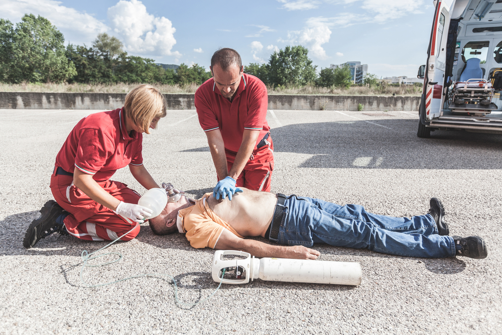
The treatment of PEA requires immediate action through high-quality cardiopulmonary resuscitation (CPR) following Advanced Cardiac Life Support (ACLS) protocols. The primary goal is to maintain vital organ perfusion while identifying and addressing the underlying cause.
For detailed guidance on performing high-quality CPR, refer to this solo provider adult BLS course.
The success of PEA treatment heavily depends on identifying and addressing the root cause. Each underlying condition requires a specific intervention:
For more information on managing specific conditions like heart attacks which may lead to PEA, you can refer to this resource.
The implementation of mechanical circulatory support devices might be necessary in specific situations:
After successful resuscitation, careful post-resuscitation management is crucial for patient recovery. This includes monitoring vital signs, ensuring adequate oxygenation and perfusion, and preparing for transfer to tertiary care if necessary. For more insights into post-resuscitation management, check out our detailed guide on transfer to tertiary care.
In conclusion, treating PEA involves a multifaceted approach that requires swift action, precise medication administration, and targeted interventions based on the underlying causes.

In addition to basic life support measures, specific medications are important in managing PEA.
Atropine is given when PEA is caused by a slow heart rate (bradycardia), especially in patients with:
The recommended atropine dosage starts at 0.5mg IV push, repeatable every 3-5 minutes up to a maximum of 3mg.
Sodium bicarbonate is used in PEA cases associated with:
The initial sodium bicarbonate dose typically ranges from 50-100 mEq, administered through IV bolus. Healthcare providers must carefully monitor blood pH and electrolyte levels during treatment.
These medications are used alongside continuous heart monitoring and assessment. The success of these treatments relies on:
Regular blood gas analysis helps guide the use of sodium bicarbonate, while continuous ECG monitoring assists in evaluating atropine's effectiveness.
PEA cardiac arrests present significant challenges with survival rates ranging from 2% to 15%. These statistics emphasize the critical nature of this condition and the need for rapid intervention.
Key factors affecting survival rates include:
Patients who experience PEA in hospital settings typically show better outcomes due to immediate access to advanced medical care. The survival rate increases when PEA converts to a shockable rhythm during resuscitation attempts.
Positive prognostic indicators:
Research indicates that survival rates improve with implementation of standardized protocols and regular training of medical personnel. Continuous monitoring and post-resuscitation care play crucial roles in long-term survival and neurological outcomes for PEA patients.
The development of new treatment strategies, such as the impact of AI on emergency cardiac care, offers hope for improving future survival rates in PEA cases by enhancing diagnosis, treatment precision, and patient outcomes through advanced data analysis and real-time decision support.
.jpg)