Every minute counts when cardiac arrest strikes. This life-threatening condition stops the heart from pumping blood effectively, cutting off vital oxygen supply to organs and tissues. Without immediate intervention, cardiac arrest can lead to permanent brain damage or death within minutes.
The survival rate for cardiac arrest patients depends heavily on rapid response and proper post-arrest care. Research shows that implementing a structured immediate post-cardiac arrest care algorithm can double - or even triple - survival rates.
Key survival factors include:
Healthcare providers trained in Advanced Cardiac Life Support (ACLS) follow specific algorithms to optimize patient outcomes. These protocols guide critical decisions during the "golden hour" - the crucial period immediately following successful resuscitation.
Understanding CPR basics and advanced life support principles is essential for medical professionals. The immediate post-cardiac arrest care algorithm provides a systematic approach to:
This guide breaks down each step of the post-cardiac arrest care algorithm, helping healthcare providers deliver optimal care when every second matters.

Cardiac arrest differs significantly from a heart attack. A heart attack occurs when blood flow to the heart is blocked, while cardiac arrest happens when the heart suddenly stops beating. Think of it as a heart attack being a "circulation" problem, while cardiac arrest is an "electrical" problem.
Signs of Cardiac Arrest:
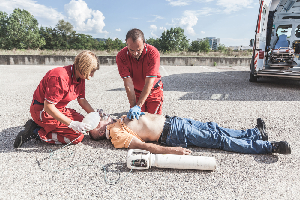
CPR serves as the critical link between cardiac arrest and survival. When performed correctly, chest compressions manually pump blood through the body, delivering vital oxygen to organs until advanced medical care arrives.
Basic Life Support (BLS) creates the foundation for successful resuscitation. BLS skills include high-quality CPR, proper use of an AED, and recognition of life-threatening emergencies. These fundamental techniques bridge the gap between cardiac arrest and advanced medical interventions, significantly increasing survival chances when performed promptly and correctly.
Healthcare providers must maintain proficiency in BLS skills through regular training and certification. This expertise enables them to respond effectively during the critical first minutes of cardiac arrest, setting the stage for successful advanced life support measures.
To ensure effective response in such scenarios, healthcare providers often undergo BLS Certification which includes comprehensive training on essential skills like high-quality CPR and proper AED usage. Regular BLS Recertification further enhances their ability to handle emergencies efficiently.
Understanding other medical emergencies like stroke can also be beneficial as these situations may require similar immediate responses and interventions.
Advanced Cardiac Life Support (ACLS) is a higher level of medical care used to treat life-threatening heart emergencies. It builds on Basic Life Support (BLS) techniques and includes more advanced procedures and medications to help patients recover.
The ACLS algorithm helps healthcare providers determine the best course of action based on the patient's condition. It outlines different pathways for treatment depending on whether the heart rhythm is shockable or non-shockable.
ACLS goes beyond basic CPR by incorporating several advanced interventions:
To provide ACLS care, healthcare providers must complete specialized training and obtain certification from courses built on American Heart Association (AHA) guidelines. These courses include:
The ACLS algorithm emphasizes the importance of delivering high-quality CPR while simultaneously identifying and addressing the underlying cause of cardiac arrest.
During training, providers learn how to:
For patients in specific age groups, like children, it's essential to adapt the ACLS protocols accordingly. This is where resources such as the Pediatric Basic Life Support Algorithm come into play, particularly in situations involving two rescuers.
Additionally, understanding child safety at home can be crucial for parents or guardians. Courses like those offered by Affordable ACLS can provide valuable insights on recognizing common household hazards and emergency response for accidents.
ACLS certification requires renewal every two years through recertification courses. These courses update providers on the latest evidence-based practices and guidelines in emergency cardiovascular care.
The impact of AI on this field is also noteworthy, as it is transforming emergency cardiac care by improving diagnosis, treatment precision, and patient outcomes through advanced data analysis and real-time decision support, as detailed in this article about the impact of AI on emergency cardiac care.
Healthcare providers seeking ACLS certification must complete specific requirements to demonstrate their competency in advanced cardiac life support and post-cardiac arrest care. Here's what you need to know about the certification process:
You'll find ACLS certification courses offered through various authorized training centers, medical facilities, and educational institutions. The American Heart Association maintains strict standards for these programs to ensure consistent quality in emergency cardiac care education. For those needing to refresh their skills, recertification courses are available that include valuable resources such as study tips and solo provider adult BLS lessons.
The immediate post-cardiac arrest care algorithm serves as a structured roadmap for healthcare providers managing patients who achieve return of spontaneous circulation (ROSC). This evidence-based protocol maximizes survival chances through systematic interventions and continuous monitoring.
ROSC marks the critical transition point between active resuscitation and post-cardiac arrest care. Healthcare providers must confirm ROSC through specific clinical indicators:
Primary ROSC Confirmation Signs:
Secondary ROSC Indicators:
Vital Signs Target Range:
Healthcare providers must document the exact time of ROSC achievement. This timestamp guides subsequent interventions and monitoring intervals. The medical team should record:
Documentation Requirements:
ROSC achievement doesn't guarantee sustained cardiac function. The medical team must maintain constant vigilance for:
Post-ROSC Monitoring:
The successful confirmation of ROSC triggers the activation of subsequent post-cardiac arrest care protocols. These interventions focus on maintaining stable cardiac function while addressing potential complications and underlying causes of the arrest.
To better understand the complexities involved in this process, including the necessary steps to ensure a successful transition from resuscitation to post-cardiac arrest care, it's essential to familiarize oneself with various scenarios that may arise. A helpful resource for this is the post-cardiac arrest care quiz, which provides valuable insights and knowledge to aid healthcare professionals in these critical situations.

After successfully restoring a patient's heartbeat (ROSC), it is crucial to secure their airway. This ensures that they can breathe properly and receive adequate oxygenation. The preferred method for managing the airway in such cases is through early endotracheal intubation.
In certain situations, it may be necessary to reposition the patient before performing intubation:
Once the airway has been secured and ventilation initiated, it is important to monitor specific parameters:
Continuous waveform capnography should be utilized for real-time assessment of ventilation:
By implementing these strategies for airway management after cardiac arrest, healthcare providers can minimize complications such as aspiration, low oxygen levels (hypoxia), and secondary injury to the brain. It is essential to remain vigilant in monitoring airway parameters while simultaneously preparing for further interventions outlined in post-arrest care protocols.
Stabilizing blood pressure remains a critical priority in post-cardiac arrest care. Your immediate goal is maintaining systolic blood pressure above 90 mm Hg or mean arterial pressure (MAP) above 65 mm Hg.
Here's your step-by-step approach to hemodynamic stabilization:
Your hemodynamic goals might need adjustment based on:
Rapid cardiac evaluation through a 12-lead ECG is a critical step in post-cardiac arrest care. This diagnostic tool helps identify acute coronary syndromes, including ST-elevation myocardial infarction (STEMI), which requires immediate intervention.
A 12-lead ECG should be obtained within 10 minutes of return of spontaneous circulation (ROSC). If STEMI is detected, immediate activation of the cardiac catheterization lab becomes necessary for potential percutaneous coronary intervention (PCI).
The cardiac evaluation process includes assessment of:
Blood samples for cardiac biomarkers should be drawn, including troponin levels, to assess the extent of myocardial injury. These markers help guide treatment decisions and provide prognostic information for the healthcare team.
Neurological assessment stands as a critical component in post-cardiac arrest care. Your immediate evaluation focuses on two key patient states:
The implementation of TTM requires precise temperature control and monitoring. Your cooling methods might include:
Sedation and neuromuscular blockade often become necessary during TTM to prevent shivering and optimize temperature control. Regular neurological assessments continue throughout the cooling and rewarming phases, with documentation of any changes in patient status.
It's important to note that a cardiac arrest may sometimes be preceded by a heart attack, characterized by symptoms such as chest tightness, nausea, sweating, shortness of breath, fatigue, pain in the arm or jaw, and pallor. Recognizing these symptoms early can be crucial for timely intervention, which may include calling emergency services or administering CPR if necessary.
Critical care monitoring is essential for managing patients after a cardiac arrest. Using specific temperature monitoring methods can significantly impact patient recovery.
To ensure effective temperature monitoring, follow these best practices:
Temperature monitoring devices should connect to central monitoring systems, allowing healthcare providers to track trends and respond promptly to variations. Your choice of monitoring method depends on patient factors, including consciousness level, existing medical devices, and specific care requirements.
The immediate post-cardiac arrest care algorithm emphasizes rapid identification and treatment of underlying causes. Healthcare providers use a systematic approach known as the 'H's and T's' to address reversible conditions.
Each cause requires specific interventions and monitoring. For example, hypovolemia demands immediate fluid resuscitation with crystalloid solutions, while tension pneumothorax might need urgent needle decompression. Healthcare providers must maintain a high index of suspicion for these conditions during post-ROSC care.
The treatment approach should be individualized based on clinical presentation, patient history, and available diagnostic information. Quick recognition of these reversible causes can significantly improve patient outcomes in the post-cardiac arrest period.
The transfer of post-cardiac arrest patients to specialized care facilities marks a critical phase in their recovery journey. Successful patient outcomes depend on careful timing and specific criteria for transfer decisions.
The receiving facility should maintain continuous communication with the transport team during transfer. Time-sensitive interventions like cardiac catheterization or specialized neurological care guide the choice of destination facility. Direct admission to cardiac or neurological intensive care units streamlines the transition to specialized care.

The immediate post-cardiac arrest care algorithm serves as a critical extension of Advanced Cardiac Life Support (ACLS) protocols. This integration creates a seamless transition from resuscitation efforts to sustained recovery care.
The integration of these protocols requires healthcare providers to maintain proficiency in both ACLS algorithms and post-arrest care guidelines. Regular training sessions often combine both elements to reinforce their interconnected nature.
Healthcare facilities typically develop standardized order sets that bridge ACLS and post-arrest care phases. These tools help ensure consistent application of both protocols while maintaining flexibility for patient-specific needs.
For those looking to enhance their skills in managing such critical situations, Affordable ACLS offers resources that can equip individuals with the life-saving skills necessary in emergencies, including PALS certification which is particularly beneficial for those working with children who may experience sudden cardiac arrest or other medical emergencies.
The Immediate Post-Cardiac Arrest Care Algorithm is a crucial tool in emergency medicine. This structured approach turns chaotic situations into organized, life-saving actions that directly affect patient outcomes.
Research shows that healthcare providers who understand and use this algorithm achieve:
Your knowledge and use of this algorithm can be the deciding factor between life and death. Each part - from achieving ROSC to managing reversible causes - forms a complete framework for optimal patient care.
The key to improving survival in cardiac arrest lies in:
Remember: This algorithm isn't just a set of rules - it's a flexible tool that adjusts to each patient's specific needs. Your dedication to mastering these steps, staying up-to-date with certifications, and regularly practicing simulations ensures you're ready when every second matters.
The journey to saving lives begins with knowledge. Use this algorithm as your foundation for providing outstanding emergency cardiac care.
.jpg)