ACLS Blogs

Bradycardia Algorithm Companion: When to Reach for the Pacer

Why the Bradycardia Algorithm Trips Up Even Experienced Providers

Tachycardia gets the dramatic energy settings. Cardiac arrest gets the compressions. Bradycardia gets... a slow, quiet drift toward a decision that many providers make too late. The ACLS bradycardia algorithm looks simple on paper, but the clinical question underneath it is not: this patient has a slow heart rate, so is the rate actually the problem, and if it is, what do I do right now?


This is a companion to the full Adult Bradycardia with a Pulse algorithm page. That page walks the flowchart box by box. Here we focus on the part the flowchart cannot show you: the decision path, the moment you stop waiting on drugs and reach for the pacer, and how to set that pacer up so it actually works. We are practicing emergency physicians who teach this at the bedside, so expect the practical version.


One note on scope. If you have read our older piece on symptomatic bradycardia causes and treatment, that article covers the broad picture of why hearts slow down. This one is provider-facing and built for the megacode station and the real resuscitation room: what to decide, in what order, and why.


The One Question That Drives the Whole Algorithm

Before any box, ask whether the slow rate is causing the patient's problem. A trained endurance athlete at 48 beats per minute who is comfortable and talking is not a bradycardia emergency. An elderly patient at 38 who is gray, confused, and hypotensive is. The algorithm only starts moving when the answer is yes.


In ACLS language, you are looking for a persistent bradyarrhythmia that is causing acute signs of instability. Those signs are the trigger for treatment:


  • Hypotension or other signs of shock, such as cool, mottled skin and delayed capillary refill
  • Acutely altered mental status, including new confusion, agitation, or drowsiness
  • Ischemic chest discomfort that fits the bradycardia timeline
  • Acute heart failure, such as new pulmonary edema or rising work of breathing


Notice what is not on that list: the number itself. Providers sometimes fixate on a rate below 50 or 60. Current AHA guidance frames treatment around hemodynamic compromise and symptoms attributable to the slow rhythm. The AHA adult advanced life support guideline says evaluation and treatment of reversible causes is recommended in acute symptomatic bradycardia, and that atropine is reasonable when bradycardia comes with hemodynamic compromise.


The Decision Path in Plain Language

Step 1: Support the patient while you think

Airway, breathing, circulation. Give oxygen if the patient is hypoxemic, attach the monitor, get IV access, and obtain a 12-lead ECG if it does not delay care. Do these in parallel with your assessment, not as a sequence. In the megacode, saying out loud that you are placing the patient on a monitor and establishing access earns you points and keeps the scenario moving.


Step 2: Hunt for the reversible cause

Bradycardia is often a symptom of something else. Ask about new medications (beta blockers, calcium channel blockers, digoxin, and other rate-slowing drugs), look for hypoxia, electrolyte problems, hypothermia, and ischemia, particularly an inferior myocardial infarction. If you can fix the cause, you may not need the pacer at all. If the patient is unstable, though, you treat the rate while you search. Do not let the diagnostic workup become a reason to delay.


Step 3: Atropine first, but briefly

For an unstable patient, atropine remains the first drug in the algorithm. In the 2020 update the first dose was 1 mg IV, repeated every 3 to 5 minutes up to a 3 mg total, so confirm the dose against the algorithm version your course teaches. We keep atropine detail short here because the point of this companion is the decision, not the drug. What matters: atropine is a bridge, not a destination, and it works poorly in some rhythms that you should learn to recognize on sight.


Step 4: Escalate to the pacer, an infusion, or both

If the bradycardia persists and the patient is still unstable, the algorithm offers two escalation options: transcutaneous pacing, or a chronotropic infusion such as dopamine or epinephrine. Both are meant to be used while you prepare for expert consultation and possible transvenous pacing. The next section is about how to choose between them, because that is where most of the clinical judgment lives.

Clinicians assessing a patient with slow heart rhythm on a cardiac monitor


When to Reach for the Pacer

The textbook sequence is atropine, then pacing or an infusion. Real patients do not always read the textbook. Here are the situations where you should be thinking about the pacer before the atropine clock runs out.


High-grade AV block with a wide QRS

Atropine acts by blocking vagal tone at the SA and AV nodes. If the block sits below the AV node, in the His-Purkinje system, there is little vagal tone to block, and atropine may be ineffective or can even worsen conduction. Second-degree Mobitz type II and third-degree block with a wide-complex escape rhythm are the classic examples. If you recognize one of these in an unstable patient, pads on and pacing prepared immediately is a defensible and often better first move. Our guide to first, second, and third degree heart blocks is the fastest way to sharpen that recognition.


Atropine has not worked

If you have given atropine and the patient remains unstable, do not keep re-dosing hoping for a different result. Move on. A patient who is still hypotensive and confused after a reasonable atropine trial needs a rate you can control.


The patient is deteriorating fast

Pre-arrest bradycardia is real. A heart rate falling toward the 20s with fading perfusion should push you to apply pads early, even while atropine goes in. Pacing takes preparation time, and that time is the one thing an unstable patient does not have. Many experienced clinicians apply the pads as the second thing they do, not the last.


Special populations where atropine is unreliable

The ACLS algorithm notes that atropine is unlikely to help in a denervated transplanted heart, because the vagus nerve no longer reaches it. Expect to move to pacing or an adrenergic infusion quickly in that setting, and involve specialist help early.


Pacer or infusion: how to choose

The AHA lists dopamine and epinephrine infusions alongside transcutaneous pacing as options after atropine. The honest clinical split looks like this:


  • Pacing is favored when block is infranodal, when the patient is profoundly unstable, when the rhythm is failing fast, or when drugs are likely to be ineffective
  • An infusion is reasonable when pacing is not immediately available, when the patient cannot tolerate pacing discomfort, or as a bridge while pads and sedation are being prepared
  • Both together is common in practice: infusion running while the pacer is set up, with one covering the gap for the other


Whichever you choose, say it out loud and say why. In a megacode, examiners are listening for your reasoning as much as the action. For drug ranges, keep our ACLS medications cheat sheet nearby, and consult the ACLS algorithms hub for the other rhythm pathways.


How to Set Up Transcutaneous Pacing Without Fumbling

Pacing is a procedure, and procedures are easier when you rehearse the steps. Our dedicated guide to transcutaneous pacing: when and how to use it goes deeper. Here is the compressed checklist, using the order that keeps you out of trouble.


  • Pads on clean, dry skin. Dry the chest, clip excess hair if needed, and avoid placing pads over medication patches or implanted devices
  • Position the pads. The classic placements are anterior-posterior or anterolateral. A recent PMC study comparing pad positions in symptomatic bradycardia is worth reading if you want to see how this is being examined
  • Select demand mode and set a rate. Demand mode lets the device pace only when the patient's own rate falls below your set rate. A rate in the range of 60 to 80 beats per minute is commonly used
  • Start the current low and increase. Raise output until you see electrical capture, a wide QRS with a pacer spike followed by a T wave
  • Confirm mechanical capture. This is the step people skip. Check for a palpable pulse, ideally at the femoral artery, and watch the blood pressure and perfusion
  • Add a safety margin and treat pain. Once capture is reliable, many protocols add a bit of output above threshold. Provide analgesia or sedation as the situation allows, because pacing is uncomfortable


The electrical capture trap

The monitor can show a beautiful wide-complex paced rhythm while the patient has no pulse. Electrical capture is not mechanical capture. If you do not feel a pulse that matches the pacer rate, you have not succeeded, and you should reassess pad position, increase output, and look for causes such as hypoxia or acidosis. A hands-on pulse check beats a monitor trace every time. Muscle twitching from the pacer can also mimic a pulse, which is another reason to confirm with a central pulse and the clinical picture.


Know the limits of the evidence too. A systematic review of prehospital transcutaneous pacing found inadequate evidence to determine its efficacy for symptomatic bradycardia in that setting, and no evidence supporting use in bradyasystolic arrest. Pacing is a reasonable bridge for the unstable patient with a pulse, not a cure and not a rescue for asystole. That is why the algorithm pairs it with expert consultation and planning for transvenous pacing.
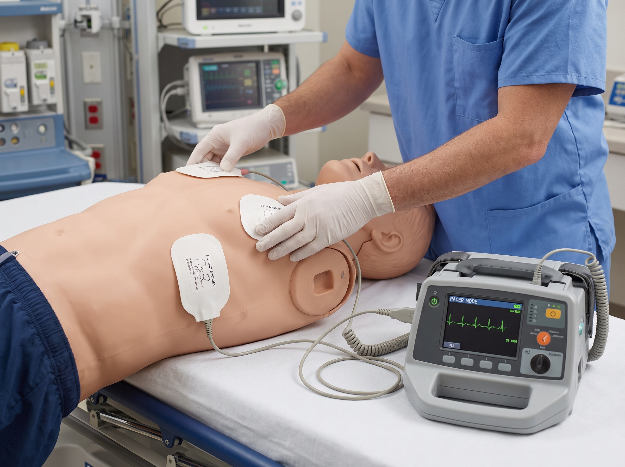
Transcutaneous pacing pad placement on a training mannequin


Megacode and Exam Notes for Bradycardia

If you are preparing for the skills station or the written exam, these are the bradycardia moments that cost points most often. Practice them out loud:


  • Treating the number, not the patient. Always state whether the patient is stable or unstable and name the signs that decided it
  • Forgetting the reversible causes. Even in an unstable patient, announce that you are looking for and treating contributing causes
  • Re-dosing atropine on a block that will not respond. Recognize infranodal block and move on to pacing
  • Skipping mechanical capture. Say that you are confirming a pulse with capture, every time
  • Mixing up the tachycardia pathway. The bradycardia and tachycardia algorithms both hinge on stability, but the interventions are different; our tachycardia algorithm companion is the matching reference for the fast side
  • Late call for help. Say early that you are requesting expert consultation and preparing for transvenous pacing if needed


The best fix is repetition under realistic pressure. Our free practice tests let you drill the stable versus unstable decision with rationales, and the older explainer Managing Bradycardia: The ACLS Algorithm Explained is a good second read if you want a different angle on the same flowchart.


What Happens After the Pacer Goes On

Pacing stabilizes the patient but does not finish the case. Once you have a reliable rate and perfusion, the plan moves to the underlying problem. Is there an ischemic cause that needs the cath lab? Is a drug effect the culprit and does it need a specific antidote or removal? Does the patient need transvenous pacing or a permanent device? The ACC/AHA/HRS guideline on bradycardia and cardiac conduction delay addresses the longer evaluation and the question of permanent pacing, which sits outside the scope of the ACLS algorithm but is where many of these patients are headed.


Document the time you applied pads, the capture threshold, the rate and output you settled on, and the patient's response. These details matter when the receiving team picks up the case, and they are the kind of closing-the-loop communication that team-dynamics sections of the ACLS exam like to see.


Finally, remember that pediatric bradycardia flips much of this logic. In children, bradycardia is more often caused by hypoxia than by primary conduction disease, and the first response is oxygenation and ventilation. If you work across age groups, our comparison of pediatric versus adult bradycardia is worth the read.


Bradycardia Quick Reference Card

Screenshot this or write it on the back of your hand before the megacode. It is a memory aid, not a replacement for your course's current algorithm.


  • Ask: Is the slow rate causing hypotension, altered mental status, ischemic chest pain, or acute heart failure?
  • If no: Monitor, find the cause, and observe
  • If yes: Support ABCs, monitor, IV access, 12-lead if it will not delay care, treat reversible causes
  • Atropine is first-line for most unstable patients, but not for infranodal block or a denervated heart
  • Still unstable: Transcutaneous pacing or dopamine/epinephrine infusion while calling for expert consultation and preparing for transvenous pacing
  • Pacing: Pads, demand mode, set the rate, raise output to capture, confirm a pulse, treat pain
  • Watch for arrest: If the pulse is lost, leave the bradycardia pathway and start the cardiac arrest algorithm


Putting It Into Practice

Bradycardia decisions are quick, but they are learnable. The pattern is consistent: judge the patient first, hunt for the cause, try atropine where it makes sense, and do not be shy about pacing when the rhythm or the trajectory tells you drugs will not be enough. If your certification is coming due or you are working toward your first card, our online ACLS course covers bradycardia, tachycardia, arrest rhythms, and the megacode in a self-paced format built by practicing emergency physicians. Retakes are unlimited at no extra charge, and the money-back guarantee applies if your employer does not accept the certification.


Come back to this companion any time you want a refresher on the decision path, and pair it with the full algorithm page when you want the flowchart itself in front of you.


ACLS Blogs

Bradycardia Algorithm Companion: When to Reach for the Pacer

The ACLS bradycardia algorithm is easy to read and hard to apply at the bedside. This companion walks through the decision path, when to move from atropine to the pacer, and how to confirm capture.

Why the Bradycardia Algorithm Trips Up Even Experienced Providers

Tachycardia gets the dramatic energy settings. Cardiac arrest gets the compressions. Bradycardia gets... a slow, quiet drift toward a decision that many providers make too late. The ACLS bradycardia algorithm looks simple on paper, but the clinical question underneath it is not: this patient has a slow heart rate, so is the rate actually the problem, and if it is, what do I do right now?


This is a companion to the full Adult Bradycardia with a Pulse algorithm page. That page walks the flowchart box by box. Here we focus on the part the flowchart cannot show you: the decision path, the moment you stop waiting on drugs and reach for the pacer, and how to set that pacer up so it actually works. We are practicing emergency physicians who teach this at the bedside, so expect the practical version.


One note on scope. If you have read our older piece on symptomatic bradycardia causes and treatment, that article covers the broad picture of why hearts slow down. This one is provider-facing and built for the megacode station and the real resuscitation room: what to decide, in what order, and why.


The One Question That Drives the Whole Algorithm

Before any box, ask whether the slow rate is causing the patient's problem. A trained endurance athlete at 48 beats per minute who is comfortable and talking is not a bradycardia emergency. An elderly patient at 38 who is gray, confused, and hypotensive is. The algorithm only starts moving when the answer is yes.


In ACLS language, you are looking for a persistent bradyarrhythmia that is causing acute signs of instability. Those signs are the trigger for treatment:


  • Hypotension or other signs of shock, such as cool, mottled skin and delayed capillary refill
  • Acutely altered mental status, including new confusion, agitation, or drowsiness
  • Ischemic chest discomfort that fits the bradycardia timeline
  • Acute heart failure, such as new pulmonary edema or rising work of breathing


Notice what is not on that list: the number itself. Providers sometimes fixate on a rate below 50 or 60. Current AHA guidance frames treatment around hemodynamic compromise and symptoms attributable to the slow rhythm. The AHA adult advanced life support guideline says evaluation and treatment of reversible causes is recommended in acute symptomatic bradycardia, and that atropine is reasonable when bradycardia comes with hemodynamic compromise.


The Decision Path in Plain Language

Step 1: Support the patient while you think

Airway, breathing, circulation. Give oxygen if the patient is hypoxemic, attach the monitor, get IV access, and obtain a 12-lead ECG if it does not delay care. Do these in parallel with your assessment, not as a sequence. In the megacode, saying out loud that you are placing the patient on a monitor and establishing access earns you points and keeps the scenario moving.


Step 2: Hunt for the reversible cause

Bradycardia is often a symptom of something else. Ask about new medications (beta blockers, calcium channel blockers, digoxin, and other rate-slowing drugs), look for hypoxia, electrolyte problems, hypothermia, and ischemia, particularly an inferior myocardial infarction. If you can fix the cause, you may not need the pacer at all. If the patient is unstable, though, you treat the rate while you search. Do not let the diagnostic workup become a reason to delay.


Step 3: Atropine first, but briefly

For an unstable patient, atropine remains the first drug in the algorithm. In the 2020 update the first dose was 1 mg IV, repeated every 3 to 5 minutes up to a 3 mg total, so confirm the dose against the algorithm version your course teaches. We keep atropine detail short here because the point of this companion is the decision, not the drug. What matters: atropine is a bridge, not a destination, and it works poorly in some rhythms that you should learn to recognize on sight.


Step 4: Escalate to the pacer, an infusion, or both

If the bradycardia persists and the patient is still unstable, the algorithm offers two escalation options: transcutaneous pacing, or a chronotropic infusion such as dopamine or epinephrine. Both are meant to be used while you prepare for expert consultation and possible transvenous pacing. The next section is about how to choose between them, because that is where most of the clinical judgment lives.

Clinicians assessing a patient with slow heart rhythm on a cardiac monitor


When to Reach for the Pacer

The textbook sequence is atropine, then pacing or an infusion. Real patients do not always read the textbook. Here are the situations where you should be thinking about the pacer before the atropine clock runs out.


High-grade AV block with a wide QRS

Atropine acts by blocking vagal tone at the SA and AV nodes. If the block sits below the AV node, in the His-Purkinje system, there is little vagal tone to block, and atropine may be ineffective or can even worsen conduction. Second-degree Mobitz type II and third-degree block with a wide-complex escape rhythm are the classic examples. If you recognize one of these in an unstable patient, pads on and pacing prepared immediately is a defensible and often better first move. Our guide to first, second, and third degree heart blocks is the fastest way to sharpen that recognition.


Atropine has not worked

If you have given atropine and the patient remains unstable, do not keep re-dosing hoping for a different result. Move on. A patient who is still hypotensive and confused after a reasonable atropine trial needs a rate you can control.


The patient is deteriorating fast

Pre-arrest bradycardia is real. A heart rate falling toward the 20s with fading perfusion should push you to apply pads early, even while atropine goes in. Pacing takes preparation time, and that time is the one thing an unstable patient does not have. Many experienced clinicians apply the pads as the second thing they do, not the last.


Special populations where atropine is unreliable

The ACLS algorithm notes that atropine is unlikely to help in a denervated transplanted heart, because the vagus nerve no longer reaches it. Expect to move to pacing or an adrenergic infusion quickly in that setting, and involve specialist help early.


Pacer or infusion: how to choose

The AHA lists dopamine and epinephrine infusions alongside transcutaneous pacing as options after atropine. The honest clinical split looks like this:


  • Pacing is favored when block is infranodal, when the patient is profoundly unstable, when the rhythm is failing fast, or when drugs are likely to be ineffective
  • An infusion is reasonable when pacing is not immediately available, when the patient cannot tolerate pacing discomfort, or as a bridge while pads and sedation are being prepared
  • Both together is common in practice: infusion running while the pacer is set up, with one covering the gap for the other


Whichever you choose, say it out loud and say why. In a megacode, examiners are listening for your reasoning as much as the action. For drug ranges, keep our ACLS medications cheat sheet nearby, and consult the ACLS algorithms hub for the other rhythm pathways.


How to Set Up Transcutaneous Pacing Without Fumbling

Pacing is a procedure, and procedures are easier when you rehearse the steps. Our dedicated guide to transcutaneous pacing: when and how to use it goes deeper. Here is the compressed checklist, using the order that keeps you out of trouble.


  • Pads on clean, dry skin. Dry the chest, clip excess hair if needed, and avoid placing pads over medication patches or implanted devices
  • Position the pads. The classic placements are anterior-posterior or anterolateral. A recent PMC study comparing pad positions in symptomatic bradycardia is worth reading if you want to see how this is being examined
  • Select demand mode and set a rate. Demand mode lets the device pace only when the patient's own rate falls below your set rate. A rate in the range of 60 to 80 beats per minute is commonly used
  • Start the current low and increase. Raise output until you see electrical capture, a wide QRS with a pacer spike followed by a T wave
  • Confirm mechanical capture. This is the step people skip. Check for a palpable pulse, ideally at the femoral artery, and watch the blood pressure and perfusion
  • Add a safety margin and treat pain. Once capture is reliable, many protocols add a bit of output above threshold. Provide analgesia or sedation as the situation allows, because pacing is uncomfortable


The electrical capture trap

The monitor can show a beautiful wide-complex paced rhythm while the patient has no pulse. Electrical capture is not mechanical capture. If you do not feel a pulse that matches the pacer rate, you have not succeeded, and you should reassess pad position, increase output, and look for causes such as hypoxia or acidosis. A hands-on pulse check beats a monitor trace every time. Muscle twitching from the pacer can also mimic a pulse, which is another reason to confirm with a central pulse and the clinical picture.


Know the limits of the evidence too. A systematic review of prehospital transcutaneous pacing found inadequate evidence to determine its efficacy for symptomatic bradycardia in that setting, and no evidence supporting use in bradyasystolic arrest. Pacing is a reasonable bridge for the unstable patient with a pulse, not a cure and not a rescue for asystole. That is why the algorithm pairs it with expert consultation and planning for transvenous pacing.

Transcutaneous pacing pad placement on a training mannequin


Megacode and Exam Notes for Bradycardia

If you are preparing for the skills station or the written exam, these are the bradycardia moments that cost points most often. Practice them out loud:


  • Treating the number, not the patient. Always state whether the patient is stable or unstable and name the signs that decided it
  • Forgetting the reversible causes. Even in an unstable patient, announce that you are looking for and treating contributing causes
  • Re-dosing atropine on a block that will not respond. Recognize infranodal block and move on to pacing
  • Skipping mechanical capture. Say that you are confirming a pulse with capture, every time
  • Mixing up the tachycardia pathway. The bradycardia and tachycardia algorithms both hinge on stability, but the interventions are different; our tachycardia algorithm companion is the matching reference for the fast side
  • Late call for help. Say early that you are requesting expert consultation and preparing for transvenous pacing if needed


The best fix is repetition under realistic pressure. Our free practice tests let you drill the stable versus unstable decision with rationales, and the older explainer Managing Bradycardia: The ACLS Algorithm Explained is a good second read if you want a different angle on the same flowchart.


What Happens After the Pacer Goes On

Pacing stabilizes the patient but does not finish the case. Once you have a reliable rate and perfusion, the plan moves to the underlying problem. Is there an ischemic cause that needs the cath lab? Is a drug effect the culprit and does it need a specific antidote or removal? Does the patient need transvenous pacing or a permanent device? The ACC/AHA/HRS guideline on bradycardia and cardiac conduction delay addresses the longer evaluation and the question of permanent pacing, which sits outside the scope of the ACLS algorithm but is where many of these patients are headed.


Document the time you applied pads, the capture threshold, the rate and output you settled on, and the patient's response. These details matter when the receiving team picks up the case, and they are the kind of closing-the-loop communication that team-dynamics sections of the ACLS exam like to see.


Finally, remember that pediatric bradycardia flips much of this logic. In children, bradycardia is more often caused by hypoxia than by primary conduction disease, and the first response is oxygenation and ventilation. If you work across age groups, our comparison of pediatric versus adult bradycardia is worth the read.


Bradycardia Quick Reference Card

Screenshot this or write it on the back of your hand before the megacode. It is a memory aid, not a replacement for your course's current algorithm.


  • Ask: Is the slow rate causing hypotension, altered mental status, ischemic chest pain, or acute heart failure?
  • If no: Monitor, find the cause, and observe
  • If yes: Support ABCs, monitor, IV access, 12-lead if it will not delay care, treat reversible causes
  • Atropine is first-line for most unstable patients, but not for infranodal block or a denervated heart
  • Still unstable: Transcutaneous pacing or dopamine/epinephrine infusion while calling for expert consultation and preparing for transvenous pacing
  • Pacing: Pads, demand mode, set the rate, raise output to capture, confirm a pulse, treat pain
  • Watch for arrest: If the pulse is lost, leave the bradycardia pathway and start the cardiac arrest algorithm


Putting It Into Practice

Bradycardia decisions are quick, but they are learnable. The pattern is consistent: judge the patient first, hunt for the cause, try atropine where it makes sense, and do not be shy about pacing when the rhythm or the trajectory tells you drugs will not be enough. If your certification is coming due or you are working toward your first card, our online ACLS course covers bradycardia, tachycardia, arrest rhythms, and the megacode in a self-paced format built by practicing emergency physicians. Retakes are unlimited at no extra charge, and the money-back guarantee applies if your employer does not accept the certification.


Come back to this companion any time you want a refresher on the decision path, and pair it with the full algorithm page when you want the flowchart itself in front of you.


blogs

More articles for you

all articles

Start Today!

Get certified at your own pace, on your schedule, from the comfort of your home.