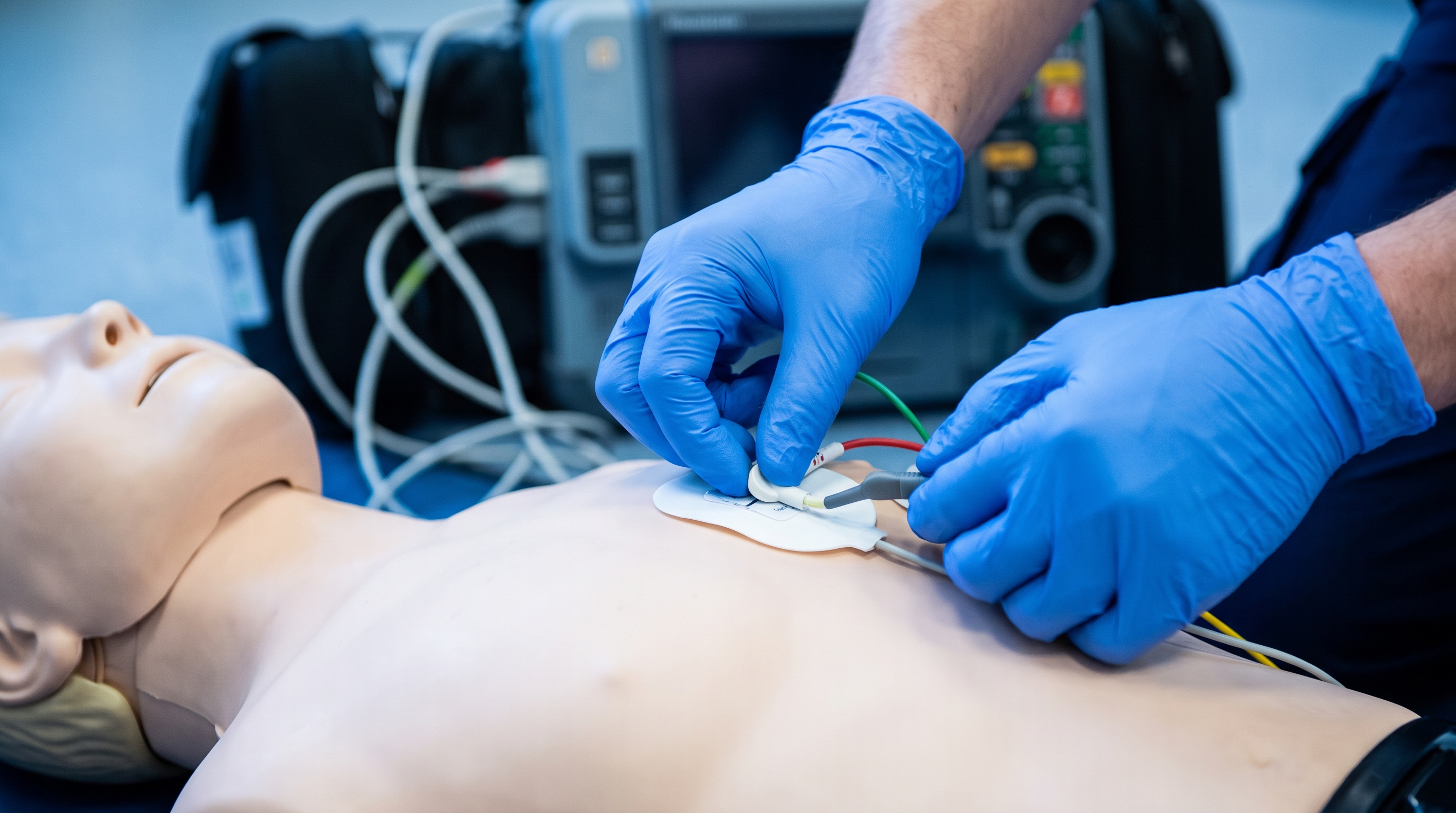
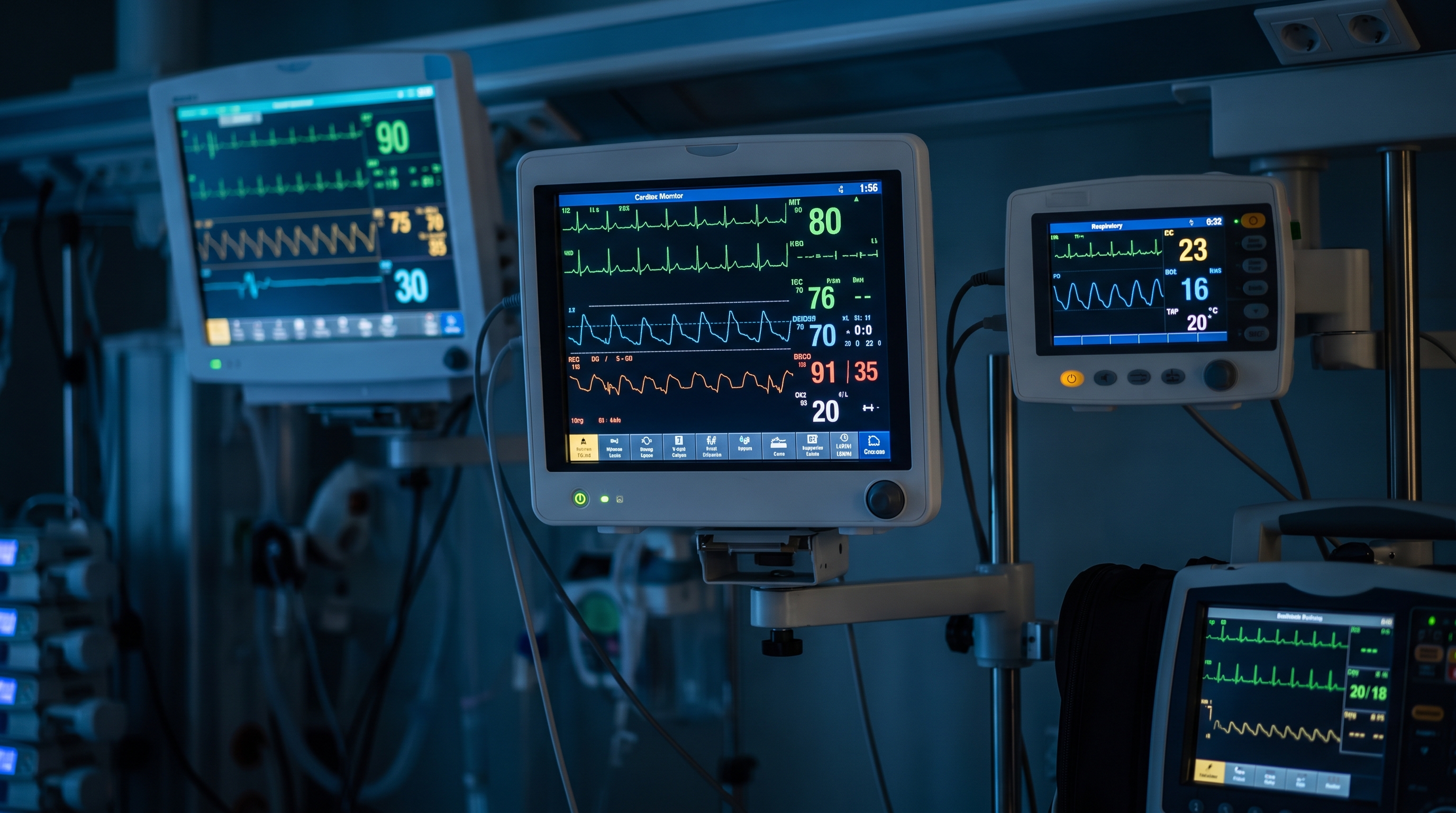
Most clinicians can recite the cardiac arrest algorithm. Far fewer can look at a four-second snippet of a monitor strip and name the rhythm before the doubt creeps in. That gap is exactly where ACLS exams and real codes get uncomfortable, because every branch of the algorithm starts with one question: what is this rhythm, and does the patient have a pulse?
This article is a self-test, not a lecture. For each of ten strips, we describe what you would see on the monitor, then stop and ask you to commit to an answer. Say it out loud or write it down. Then read the reveal, where we show the identifying criteria and the first ACLS action that follows. If you want a printable reference to keep beside you, our ACLS rhythm recognition cheat sheet covers the full set at a glance. If you are brand new to monitors, start with the beginner's guide to the core ACLS heart rhythms and come back here when the basics feel familiar.
A quick note on format: the illustrations in this article are stylized artwork, not real patient tracings. Each strip is therefore described in words, which is honestly a better test anyway. In a real code you are matching features (rate, regularity, width, P waves) rather than memorizing a picture.
Before you start, adopt the same five-question scan on every strip. It takes about ten seconds once it becomes habit, and it mirrors the approach described in the clinical literature on using a systematic approach to interpret rhythms on cardiac monitors.
Then add the question the strip cannot answer: does the patient have a pulse, and are they stable? A beautiful organized rhythm in a pulseless patient is managed very differently from the same rhythm in someone who is awake and talking. Keep that in mind as we go. Score yourself honestly; there is no penalty for missing one, and the misses tell you what to study.
A regular rhythm. Every QRS is narrow and preceded by an upright P wave, with the same distance each time. The R-to-R spacing is even but wide, with visibly more room between beats than you would expect on a normal tracing. The patient is lightheaded and the monitor shows a pressure lower than their baseline. What is your answer?
Answer: sinus bradycardia. The identifying criteria are a rate under 60, a P wave before every QRS, and a narrow complex. The ACLS question is not the name but the symptoms. If the patient is stable and asymptomatic, you observe. If there are signs of poor perfusion, you move into the bradycardia pathway, where atropine is the first drug and pacing or an infusion follows if it fails. Our bradycardia algorithm explainer walks through that decision tree.
The tracing is very fast and metronome-regular. The QRS complexes are narrow. You hunt for P waves and cannot find any you trust; at this speed they are buried in the preceding T waves. The patient says their chest is pounding. What is your answer?
Answer: supraventricular tachycardia (SVT). Think of it as narrow, regular, very fast, with no clear P waves, usually well above 150. The ACLS split is stable versus unstable. A stable patient gets vagal maneuvers and then adenosine; an unstable patient gets synchronized cardioversion. The SVT treatment decision card lays out that ladder in one page.
Narrow QRS complexes, but the spacing between them has no pattern at all. Short gap, long gap, medium gap, with no repeating sequence. The baseline between beats wobbles instead of showing distinct P waves. What is your answer?
Answer: atrial fibrillation. The two features that carry the diagnosis are an irregularly irregular rhythm and no organized P waves. A trap here is that atrial fibrillation with a rapid ventricular response can look regular at a glance when the rate climbs, so always check for those uneven gaps. In ACLS terms, you again ask whether the patient is stable and whether the fast rate is the cause of their instability or a response to something else.
A fast narrow-complex rhythm, but now the baseline has a repeating ramp-like pattern, rising and falling like the edge of a saw. The QRS complexes appear at an even rhythm, landing on every second or third ramp. What is your answer?
Answer: atrial flutter. The signature is the sawtooth flutter waves, with an atrial rate near 300 and the ventricle conducting only a fraction of them. A fixed 2:1 block yields a ventricular rate near 150, which is why flutter is a classic SVT look-alike. If you ever see a regular narrow tachycardia stuck at almost exactly 150, look hard at the baseline for flutter waves. Slowing the ventricular response briefly, for example with a vagal maneuver or adenosine, can unmask them.
This time the QRS complexes are wide and bizarre, noticeably taller and broader than a normal complex, and they march across the screen at a fast, steady rate. There are no P waves you can identify. The patient is awake but gray and sweating. What is your answer?
Answer: monomorphic ventricular tachycardia. Wide, regular, fast, uniform complexes. The word monomorphic means every complex looks like the last one. The critical next question is the pulse. With a pulse and instability, this is a synchronized cardioversion situation. Without a pulse, it is a shockable arrest rhythm and you treat it exactly like ventricular fibrillation. For the nuances of wide complex rhythms, see our guide to wide complex tachycardia myths and facts.

If you named all five correctly, the stable rhythms are in good shape. If you hesitated between SVT and flutter, or between sinus tachycardia and SVT, that is normal; those pairs trip up experienced providers too. Write down the ones you missed, because the second half of the quiz moves into the rhythms where speed of recognition changes what you do in the next ten seconds.
Wide QRS complexes at a fast rate, but unlike strip 5 they are not uniform. The amplitude swells and shrinks in waves, so the complexes appear to twist around the baseline, like a ribbon being turned. Earlier in the shift, this patient's tracing showed a long QT interval, and they had recently started a new medication. What is your answer?
Answer: torsades de pointes. This is a polymorphic ventricular tachycardia tied to a prolonged QT interval, and it can stop on its own or degenerate into ventricular fibrillation. Drug-related QT prolongation is a common culprit, as the StatPearls review of torsades de pointes explains. The ACLS emphasis is that torsades in an unstable or pulseless patient is shocked, while magnesium is the drug classically associated with it. Our clinical overview of torsades de pointes goes deeper.
There are no discernible QRS complexes, no P waves, and no regular pattern. The baseline is an irregular, quivering scribble of varying height. The patient is unresponsive and not breathing normally. What is your answer?
Answer: ventricular fibrillation (VF). It is disorganized electrical activity with no coordinated ventricular contraction, so there is no pulse. This is a shockable rhythm, and the AHA guidelines page for adult advanced life support stresses early defibrillation paired with high-quality CPR for VF and pulseless VT. Coarse VF has bigger waves and fine VF has smaller ones, but both get the same treatment. Review the family of rhythms in our article on shockable rhythms.
The monitor shows an almost flat line with a faint drift. No complexes at all. The patient is unresponsive and pulseless. Before you answer, what do you check first?
Answer: asystole, but only after you rule out a technical problem. Check that the leads are attached, the gain is turned up, and the rhythm looks flat in a second lead. A loose lead can mimic a flat line, and very fine VF can look like asystole at low gain. True asystole is a non-shockable rhythm managed with continuous high-quality CPR, epinephrine, and a search for reversible causes, which is where the Hs and Ts come in. To separate it from its non-shockable cousin, read asystole versus PEA. PEA, by the way, is the one arrest rhythm defined by the absence of a pulse rather than by how it looks, so no single strip can prove it.
Narrow QRS complexes, with regular P waves marching along at their own steady pace. The PR interval is the same on every conducted beat, with no gradual lengthening. Then suddenly a P wave appears with no QRS after it, and the pattern resumes. What is your answer?
Answer: second-degree AV block, Mobitz type II. The key is the fixed PR interval before the dropped beat. In Mobitz I the PR stretches progressively before a beat is dropped; here it does not, which is why Mobitz II is the more worrying pattern. It can progress abruptly to complete heart block, so providers plan for pacing readiness rather than relying on atropine alone. Our heart blocks made simple guide compares all three degrees side by side.
You can march out the P waves with a pair of calipers and they are regular. You can march out the QRS complexes and they are also regular, but at a much slower rate. The two never line up; the P waves wander through the QRS complexes and sometimes hide inside them. What is your answer?
Answer: third-degree (complete) AV block. The defining feature is AV dissociation: more P waves than QRS complexes, with no consistent relationship between them. As this clinical review of third-degree atrioventricular block notes, the atria and ventricles are completely disconnected. In a symptomatic patient, this is a bradycardia-pathway problem where transcutaneous pacing is often the key intervention, because atropine frequently fails when the block sits below the AV node.
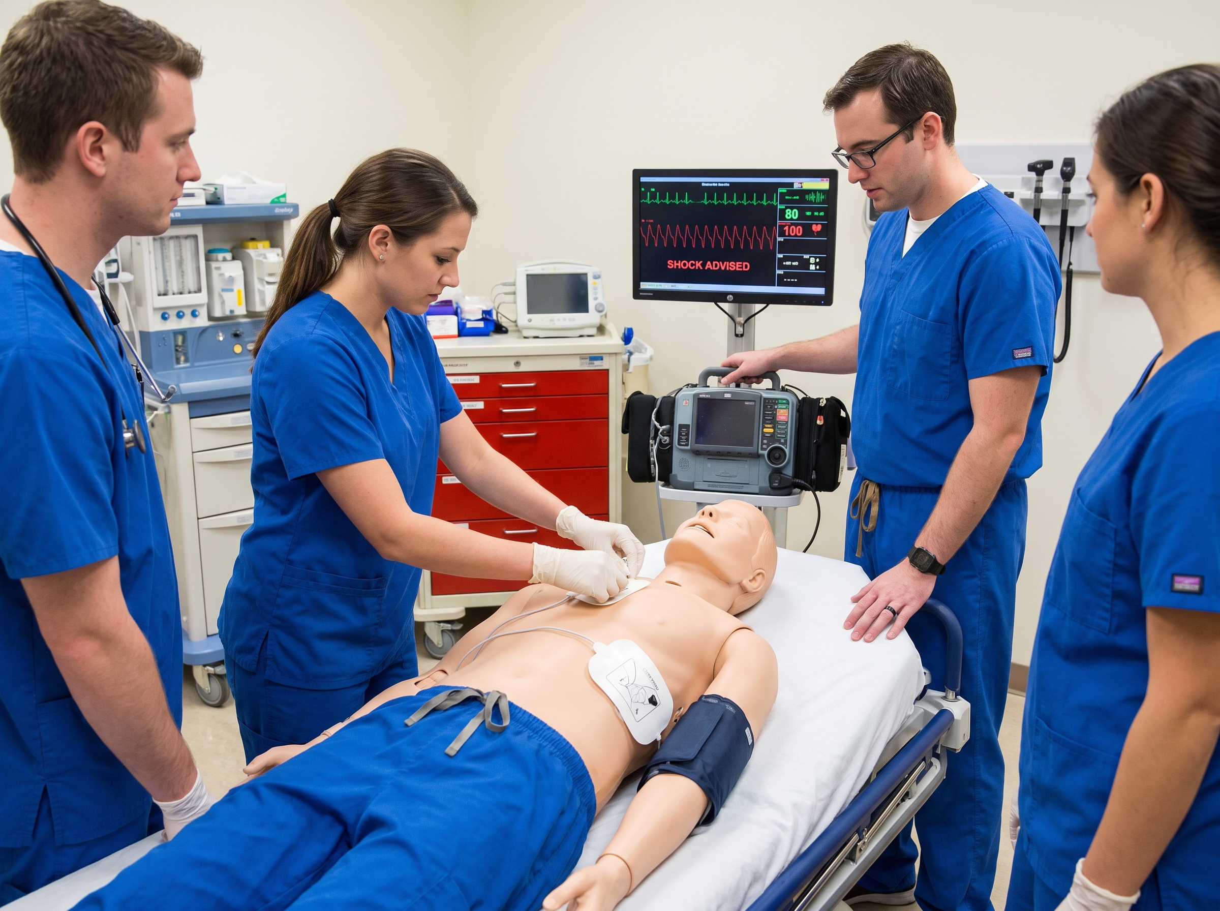
Count how many you named correctly before reading each reveal. Then use these rough bands to decide where to spend your study time:
Notice that the rhythms people miss tend to come in pairs. SVT and flutter share a regular narrow fast pattern. VF and asystole can both look flat at the wrong gain. Mobitz II and third-degree block both involve P waves that fail to conduct. Learning the pair, and the single feature that separates them, is more efficient than memorizing ten isolated pictures.
The ACLS exam rewards the provider who names the rhythm calmly, states the pulse status, and chooses the correct branch. To see these rhythms in context, walk through the five practice megacode scenarios after you finish this quiz.
Rhythm recognition is one of the few ACLS skills that fades fast without use, and it is also one of the most testable. A good routine is a short daily drill: pick two strips, run the five-question scan, and name the first action. Within a couple of weeks the scan becomes automatic. Our free practice tests are a convenient way to test the same skill under exam conditions.
When you are ready to certify or renew, Affordable ACLS online certification and recertification was built by practicing Board Certified Emergency Medicine physicians. The courses are self-paced, based on AHA and ILCOR guidelines, and include unlimited retakes and a money-back guarantee if your employer does not accept the certification, so you can study the rhythms at your own speed. Whether you work on a telemetry floor, in an emergency department, or in an ambulance, the ten strips above are the foundation. Know them cold, and the algorithms stop feeling like homework and start feeling like a plan.
.jpg)