Cardiac cath lab activation for subtle acute inferior STEMI is one of the most difficult decisions in emergency cardiology. When you come across a patient with suspected heart ischemia, the difference between timely intervention and delayed recognition can mean the difference between life and death. Subtle acute inferior STEMI cases often present without the classic ECG findings that typically trigger immediate cardiac cath lab activation, creating a diagnostic dilemma that tests even experienced clinicians.
The traditional method for activating the cath lab relies heavily on clear ST-elevation criteria visible on ECG leads. However, this standard approach misses about 55% of actual myocardial infarctions because subtle presentations don't always meet strict millimeter thresholds. You might see borderline changes in inferior leads or isolated reciprocal depression that doesn't immediately indicate the need to activate the heart cath lab.
These diagnostic challenges have serious consequences. Patients with subtle inferior STEMI are at risk of cardiac arrest and adverse outcomes just like those with obvious presentations, but they are more likely to experience treatment delays. Activating the cardiology cath team becomes a judgment call that requires integrating clinical symptoms with nuanced ECG interpretation.
In such critical situations, knowing the Adult Chain of Survival can be crucial. Early identification and appropriate activation of the cath lab can significantly improve outcomes in patients with subtle acute inferior STEMI. It's important to understand that subtle doesn't mean less dangerous – it simply means more difficult to detect.
Furthermore, once a cardiac arrest happens, following the Post Cardiac Arrest Algorithm can provide essential guidance for critical situations. Mastering these algorithms and understanding how to apply them can equip healthcare providers with vital skills to enhance patient survival rates during emergencies.
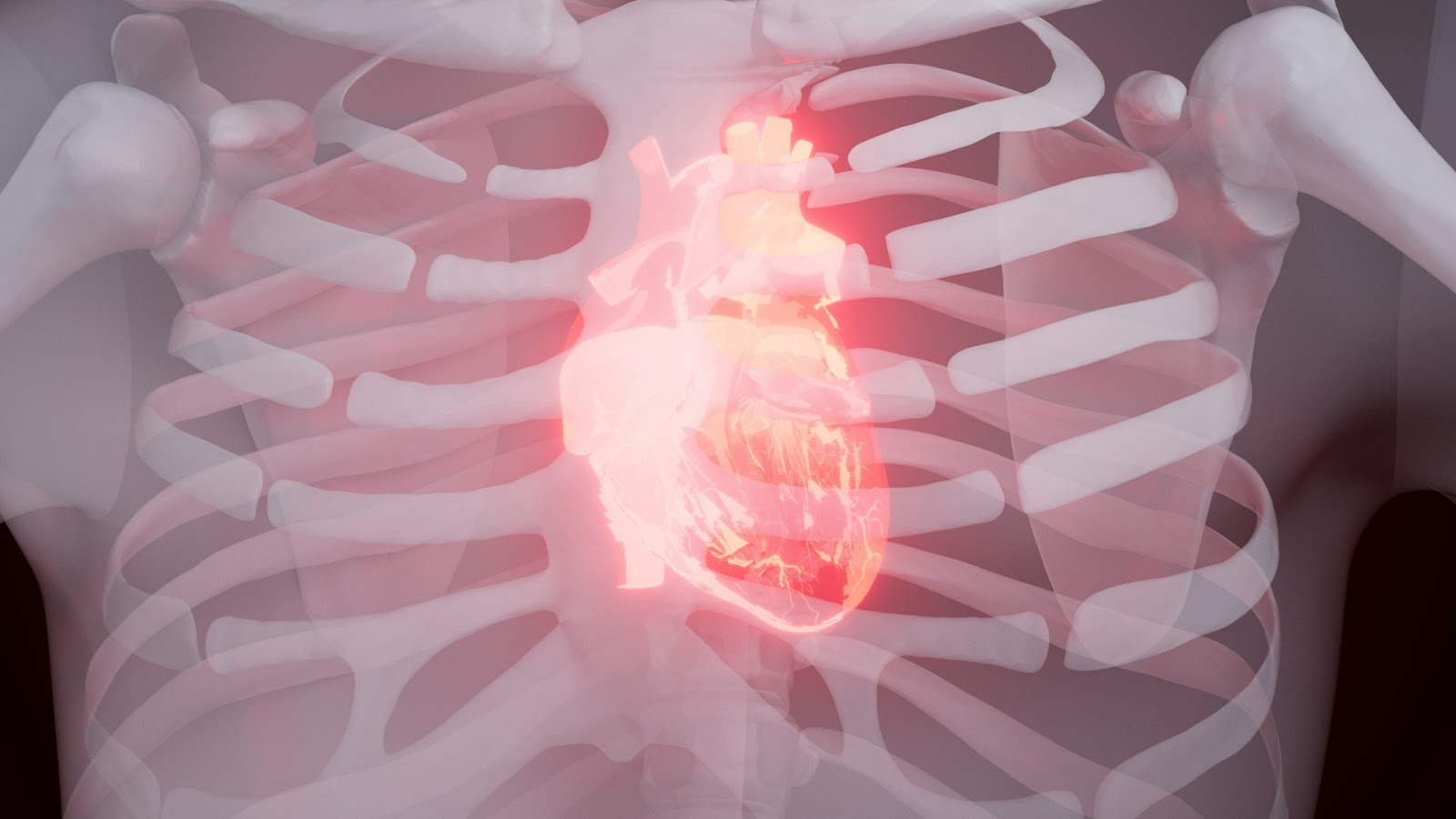
Inferior MI occurs when blood flow becomes compromised in the coronary arteries supplying the inferior wall of the left ventricle. The right coronary artery (RCA) serves as the primary culprit vessel in approximately 80-90% of cases, while the left circumflex artery accounts for the remaining 10-20%. This anatomical distribution directly impacts how ST-segment elevation myocardial infarction manifests on your heart ECG.
The inferior wall encompasses the diaphragmatic surface of the left ventricle, and when ischemia develops in this region, you'll observe characteristic changes in specific ECG leads. The pathophysiology involves complete or near-complete occlusion of the responsible coronary vessel, leading to transmural ischemia and subsequent myocardial cell death if reperfusion doesn't occur promptly.
Heart pain in inferior STEMI typically presents as:
You should recognize that inferior STEMI patients often experience more pronounced gastrointestinal symptoms compared to anterior wall infarctions. This occurs because the inferior wall's proximity to the diaphragm can trigger vagal responses, making diagnosis more challenging when patients present with primarily abdominal complaints.
It's essential to note that such emergencies can happen anywhere, including at home. Therefore, understanding child safety at home is crucial for parents or guardians. Familiarizing oneself with common household hazards and knowing how to respond during accidents can significantly improve emergency outcomes.
The 12-lead ECG serves as your primary diagnostic tool for identifying inferior STEMI. You need to focus on these specific lead changes:
Primary Leads (Inferior Territory):
Reciprocal Changes:
In addition to recognizing these signs on an ECG, it's also beneficial for medical professionals to enhance their skills through courses like BLS certification or advanced training such as ACLS algorithms. These certifications provide essential knowledge and skills that can be lifesaving in critical situations.
The current STEMI ECG criteria established by major cardiology guidelines rely on specific millimeter measurements of ST-segment elevation at the J-point. The standard thresholds require:
These ST-segment elevation criteria form the backbone of most emergency department protocols and automated ECG interpretation systems. You'll find these measurements consistently applied across hospitals worldwide, creating a standardized approach to STEMI recognition.
However, the rigid adherence to these millimeter-based thresholds creates significant diagnostic gaps. Research demonstrates that strict application of these criteria achieves only 45% sensitivity for detecting acute myocardial infarction. This means more than half of actual heart attacks may not meet the traditional STEMI definition on initial presentation.
In such cases, understanding how to manage other critical conditions like adult tachycardia, which can sometimes present alongside myocardial infarction, becomes essential. The 12 lead ECG interpretation also becomes particularly challenging when dealing with inferior wall infarctions. The inferior leads (II, III, aVF) often display subtle changes that fall just below the established thresholds. You might encounter cases where lead III shows 0.8 mm of ST elevation - technically insufficient for STEMI diagnosis yet clinically significant.
The sensitivity limitations become more pronounced in several scenarios:
These measurement-dependent criteria also fail to account for the dynamic nature of coronary occlusion. You may observe an ECG that appears normal at triage but evolves into clear STEMI patterns within 30-60 minutes. The binary nature of current guidelines - either meeting criteria or not - doesn't capture this evolving pathophysiology.
The reliance on automated ECG interpretation compounds these limitations. Computer algorithms strictly apply millimeter cutoffs without considering clinical context, potentially dismissing cases that experienced clinicians would recognize as concerning.
In addition to these challenges, it's crucial for healthcare professionals to stay updated with their certifications through recertification courses, which often include valuable insights into handling such complex scenarios effectively. Furthermore, engaging in quizzes and review lessons can significantly enhance one's understanding and application of these critical care protocols.
When standard STEMI criteria fall short, you need to develop a keen eye for subtle ECG patterns that signal acute inferior myocardial infarction. Reciprocal changes serve as your diagnostic compass in these challenging cases, providing crucial evidence of ongoing coronary occlusion even when ST elevation appears minimal or borderline.
Reciprocal changes represent the electrical mirror image of acute ischemia occurring on the opposite side of the heart. In inferior STEMI, you'll observe ST-segment depression in leads that face away from the affected territory. This phenomenon occurs because the electrical vector of injury current creates opposing deflections in leads positioned at different angles around the heart.
Lead aVL stands as the most reliable reciprocal indicator for inferior STEMI. Isolated ST depression in lead aVL demonstrates remarkable sensitivity for detecting acute inferior wall ischemia, often appearing before visible ST elevation develops in the inferior leads. You should consider any ST depression ≥0.5 mm in lead aVL as highly suspicious for inferior STEMI, particularly when accompanied by appropriate clinical symptoms.
Your diagnostic arsenal should include recognition of these specific patterns:
STEMI ECG patterns evolve rapidly, making serial monitoring essential. You might encounter a patient whose initial ECG shows only subtle ST depression in lead aVL, but subsequent tracings reveal progressive ST elevation in leads II, III, and aVF. This evolution confirms your initial suspicion and validates early intervention decisions.
The combination of reciprocal ST-segment depression with borderline inferior changes creates a complex clinical scenario that necessitates immediate action. In such cases, it's vital to follow established guideline changes for managing these patients effectively.
Furthermore, understanding and implementing proper BLS techniques during this critical period can significantly enhance patient outcomes.
Once immediate care has been administered, ensuring smooth post-resuscitation management and transfer to tertiary care is crucial for continued recovery and treatment.
When ECG findings don't fully meet traditional STEMI criteria, your clinical assessment becomes crucial in deciding between life-saving intervention and potentially fatal delays. Subtle inferior STEMI presentations require a thorough approach that considers clinical symptoms alongside borderline electrocardiographic changes.
In these challenging cases, monitoring vital signs is not enough. You need to go beyond basic monitoring and understand what the vital signs are telling you. Hypotension, especially when combined with bradycardia, strongly suggests right ventricular involvement in inferior STEMI. It's common to see patients with systolic blood pressure below 90 mmHg and heart rates under 60 beats per minute - this combination significantly increases the risk of cardiac arrest even when ST elevations appear minimal.
To integrate all this information, you need to systematically evaluate multiple clinical parameters:
It's crucial to understand that a heart attack can be characterized by one or more of the following symptoms: chest tightness, nausea, sweating, shortness of breath, fatigue, pain in the arm or jaw, and pallor. Recognizing these symptoms is vital as it can dictate immediate action such as calling 911 or preparing to start CPR if necessary.
Your clinical judgment becomes particularly important when patients present with isolated ST depression in lead aVL alongside typical ischemic symptoms. This finding, even without obvious inferior lead elevation, may represent the earliest manifestation of acute inferior STEMI requiring urgent intervention.
The risk of cardiac arrest increases rapidly in patients with subtle presentations because the underlying mechanisms are similar to classic STEMI cases. The right coronary artery blockage causing subtle ECG changes can worsen into complete vessel closure within minutes, resulting in sudden cardiac death or cardiogenic shock.
You must realize that hemodynamic compromise often occurs before clear ECG changes in inferior STEMI. Patients showing signs of right heart failure, such as elevated jugular venous pressure and swelling in their legs or ankles (peripheral edema), need immediate consideration for activating the catheterization laboratory regardless of borderline electrocardiographic findings.
In such scenarios where conventional markers fail to provide clarity, understanding the subtle indicators that suggest an impending cardiac event becomes paramount. These indicators could range from slight changes in vital signs to atypical presentations in ECG readings which should

Traditional cath lab activation protocols rely heavily on established STEMI criteria that demand specific ST-elevation thresholds measured at the J-point. These cardiology cath lab guidelines typically require ≥ 2 mm elevation for men or ≥ 1.5 mm for women in leads V2-V3, and ≥ 1 mm in other contiguous leads. While these standardized protocols work effectively for obvious cases, they create significant gaps when dealing with subtle presentations.
The rigid adherence to millimeter-based criteria poses substantial limitations in catheter lab activation decisions. Research demonstrates that strict application of these thresholds misses approximately 55% of acute myocardial infarctions, particularly those presenting with borderline or evolving changes. You face a clinical dilemma when patients present with compelling symptoms but fail to meet traditional ECG benchmarks.
Standard lab cath activation protocols often exclude patients with:
Modern cath lab activation protocols require evolution to address these diagnostic challenges. Healthcare systems need comprehensive educational initiatives that train clinicians to recognize subtle ECG patterns beyond traditional criteria. These adapted protocols should incorporate:
Cardiac Cath Lab Activation for Subtle Acute Inferior STEMI requires careful consideration of both maximizing patient outcomes and minimizing unnecessary interventions. Understanding both sides of this equation helps you make informed decisions when facing ambiguous clinical scenarios.
The early revascularization advantages become clear when you think about how important time is in preventing heart tissue damage. Every minute that passes without restoring blood flow leads to more heart tissue dying, so it's crucial to act quickly for the best results.
Key benefits include:
You need to carefully consider the risks associated with heart catheterization, especially when ECG findings are unclear or subjective interpretation influences decision-making. Activating the cath lab unnecessarily can expose patients to avoidable dangers during the procedure.
Potential complications include:
The challenge is to differentiate true subtle STEMI from other conditions that may appear similar on an ECG. When deciding whether to activate the cath lab, you should take into account factors specific to each patient, their clinical presentation, and any changes seen in serial ECG readings rather than solely relying on initial borderline findings.
In some cases, it may be necessary to move the patient to ensure their safety or provide essential care. However, moving a victim is generally not recommended unless there is a direct danger to their life. In such situations, it's crucial to assist them into a recovery position if they're unconscious but still breathing and have a pulse. This position protects their airway and minimizes the risk of aspiration. For more detailed guidance on this aspect, refer to this resource on moving victims.
Additionally, it's worth noting that there are significant benefits associated with early activation of the cath lab which can lead to improved patient outcomes when dealing with acute inferior STEMI cases.
A 58-year-old male presented to the emergency department with chest discomfort lasting 45 minutes. His initial 12-lead ECG showed minimal ST elevation in lead III (0.8 mm) with subtle ST depression in lead aVL (0.5 mm). The ECG failed to meet traditional STEMI ECG interpretation criteria, yet the attending cardiologist noted the patient's diaphoresis, nausea, and ongoing chest pain despite nitroglycerin administration.
The cardiologist's clinical judgment examples proved crucial when she activated the cath lab based on the constellation of symptoms and reciprocal changes. A repeat ECG performed 20 minutes later revealed progressive ST elevation in leads II, III, and aVF (2.1 mm, 2.8 mm, and 1.9 mm respectively) with deepening reciprocal depression in leads I and aVL. Cardiac catheterization revealed a 99% occlusion of the right coronary artery, confirming acute inferior STEMI.
A 62-year-old woman arrived with atypical chest pressure and shortness of breath. Her presenting ECG demonstrated isolated ST depression in lead aVL (1.2 mm) without clear ST elevation in inferior leads. The emergency physician initially considered this non-specific, but the patient's persistent symptoms and elevated troponin levels prompted consultation with cardiology.
The consulting cardiologist recognized that isolated ST depression in aVL can be highly sensitive for acute inferior STEMI. Serial ECGs performed every 15 minutes showed:
This dynamic evolution confirmed the initial clinical suspicion, leading to successful primary percutaneous coronary intervention.
In such critical scenarios, having well-established protocols like ACLS algorithms can significantly enhance patient outcomes by providing structured guidance for emergency care training and improving life-saving skills effectively.
Additionally, understanding specialized medical procedures such as the Pediatric Basic Life Support Algorithm, especially when dealing with pediatric patients during emergencies, is essential for ensuring optimal care and survival rates.
The importance of 12 lead ecg monitoring becomes paramount when dealing with subtle acute inferior STEMI presentations that may not immediately reveal diagnostic changes. Ischemic patterns evolve dynamically, and what appears non-diagnostic on the initial ECG can transform into clear STEMI criteria within minutes to hours.
To effectively capture these dynamic changes, serial ECG monitoring should be implemented at 15-30 minute intervals during the first few hours of patient presentation. This approach captures the progressive nature of coronary occlusion as it develops from partial to complete vessel blockage. Many subtle inferior STEMIs begin with minimal ST elevation in lead III that gradually becomes more pronounced, while reciprocal depression in leads I and aVL intensifies over time.
Serial monitoring reveals several critical progression patterns:
However, there are instances where ECG findings remain ambiguous. In such cases, the advanced cardiac imaging complementary use enhances diagnostic accuracy. Bedside echocardiography can identify new wall motion abnormalities in the inferior segments before ECG changes become diagnostic. Point-of-care ultrasound can also detect regional wall motion abnormalities that correlate with the suspected vascular territory.
Moreover, cardiac biomarkers complement serial ECG monitoring by providing biochemical evidence of myocardial injury. Troponin levels typically rise 3-6 hours after symptom onset, but high-sensitivity assays can detect elevation within 1-2 hours. Therefore, troponin measurements should be obtained every 6-8 hours when ECG findings remain subtle but clinical suspicion remains high.
The combination of serial ECG monitoring with advanced diagnostics creates a comprehensive surveillance strategy that significantly improves detection rates of evolving inferior STEMI cases that might otherwise go unnoticed.
Cardiac cath lab guidelines must evolve beyond rigid millimeter criteria to incorporate clinical judgment and subtle ECG findings. You need to develop a systematic approach that combines multiple diagnostic elements for optimal subtle acute inferior STEMI recognition.
Cardiac Cath Lab Activation for Subtle Acute Inferior STEMI requires immediate transport to PCI-capable centers, even when ECG findings remain borderline. You should prioritize facilities with 24/7 interventional capabilities and experienced teams familiar with subtle presentations.
Effective management depends on collaborative assessment between emergency medicine physicians and interventional cardiologists. You must establish protocols that allow for rapid consultation and shared decision-making when ECG findings are ambiguous but clinical suspicion remains high.
Early recognition through integrated clinical assessment and nuanced ECG interpretation significantly improves patient outcomes in subtle inferior STEMI cases.
Furthermore, enhancing your understanding of the subtle nuances in ECG interpretation can be greatly beneficial. To aid in this process, consider exploring some best study tips tailored for online course takers which could help you excel in your studies and succeed in your certification journey.
.jpg)