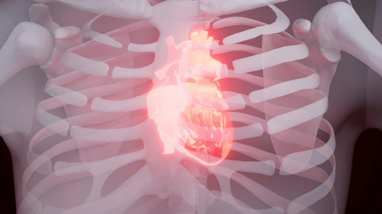
Acute Inferior STEMI with Right Ventricular Infarction and Cardiac Arrest represents one of the most challenging cardiovascular emergencies you'll encounter in clinical practice. This complex condition occurs when the right coronary artery becomes blocked, causing damage to both the inferior wall of the left ventricle and the right ventricle simultaneously. The clinical significance cannot be overstated - this combination affects approximately 50% of acute inferior STEMI cases and carries substantially higher morbidity and mortality rates compared to isolated left ventricular infarction.
Cardiac arrest in the context of myocardial infarction transforms an already serious condition into a life-threatening emergency requiring immediate intervention. When myocardial arrest occurs during acute inferior STEMI with right ventricular involvement, you're dealing with compromised cardiac output from multiple mechanisms - ischemic damage, arrhythmias, and hemodynamic instability. The right ventricular infarction creates unique challenges including elevated right-sided pressures, reduced left ventricular preload, and potential for complete cardiovascular collapse.
In such critical situations, understanding the adult chain of survival is essential. This framework guides healthcare providers in delivering effective care during a cardiac emergency.
This comprehensive guide will equip you with essential knowledge about:
Understanding these critical concepts will enhance your ability to recognize, diagnose, and manage patients facing this cardiovascular emergency. Furthermore, mastering skills related to post-cardiac arrest management is crucial for improving patient outcomes in these high-stakes scenarios.
Acute inferior STEMI occurs when the right coronary artery (RCA) is completely blocked, usually due to the rupture of an atherosclerotic plaque followed by the formation of a blood clot. The RCA is responsible for supplying blood to the inferior wall of the left ventricle, the posterior wall, and in most patients (85-90%), the right ventricle through its branches called the posterior descending artery and right ventricular marginal branches.
The right coronary artery has a specific anatomical pathway that makes it more prone to certain types of blockages. When there is a blockage in the proximal part of the RCA, it affects multiple areas of the heart at once:
This involvement of multiple territories explains why acute coronary syndrome affecting the RCA can lead to more complex clinical presentations compared to other coronary arteries.
Inferior myocardial infarction differs significantly from anterior or lateral MIs in several important aspects:
In such complex clinical situations, it is crucial for healthcare providers to be adequately prepared. This is where advanced training like BLS Certification or PALS courses could make a significant difference. These programs equip medical professionals with essential skills to handle emergencies effectively, including those associated with acute coronary syndromes.

Right ventricular infarction complicates approximately 50% of acute inferior STEMI cases, transforming what might be a manageable cardiac event into a life-threatening emergency. This high prevalence makes RVI recognition a critical skill for healthcare providers managing patients with inferior infarct patterns on their heart ECG.
The complications associated with RVI extend far beyond simple myocardial damage. When the right ventricle loses its contractile function, you're dealing with a cascade of hemodynamic problems that can rapidly progress to cardiac arrest. The compromised right ventricular function creates a domino effect throughout the cardiovascular system, affecting both cardiac output and systemic perfusion.
The hemodynamic profile of RVI creates a unique clinical picture that differs significantly from isolated left ventricular infarctions. You'll observe:
These hemodynamic changes explain why patients with RVI often present with hypotension despite having clear lung fields - a paradox that can confuse clinicians unfamiliar with heart infarction symptoms specific to right ventricular involvement.
Recognizing RVI requires you to look beyond typical chest pain presentations. The clinical syndrome includes several distinctive features:
Physical examination findings:
Given the potential for these symptoms to escalate into more severe conditions such as cardiac arrest, it's crucial for healthcare providers to be adept at recognizing and managing these signs. In such scenarios, following established protocols like the Adult Tachycardia with a Pulse Algorithm can be invaluable. Additionally, continuous education through recertification courses and self-assessment quizzes can further enhance a provider's readiness to handle these critical situations effectively.
The 12 lead ECG is the primary tool used to diagnose acute inferior STEMI with right ventricular involvement. It's important to identify specific patterns that set this complex condition apart from isolated left ventricular infarction.
ST-segment elevation in the inferior leads provides the initial diagnostic clue. Characteristic changes will be observed in:
The magnitude of ST elevation in lead III typically exceeds that in lead II when right ventricular involvement is present. This pattern helps differentiate true inferior STEMI from other causes of ST changes.
The V4R lead becomes essential for confirming right ventricular infarction. Right-sided chest leads, particularly V4R, which is placed in the fifth intercostal space at the right midclavicular line, must be obtained.
ST elevation ≥1mm in V4R is pathognomonic for right ventricular involvement and significantly impacts treatment approach. This finding occurs in approximately 50% of inferior STEMI cases and indicates a higher risk profile.
You should also evaluate for:
The combination of inferior ST elevation with V4R changes creates a diagnostic signature for Acute Inferior STEMI with Right Ventricular Infarction and Cardiac Arrest risk. You must obtain the ECG within 10 minutes of patient arrival, as early recognition directly correlates with improved outcomes through timely intervention strategies.
In addition to these diagnostic measures, it's vital to remember the importance of post-resuscitation management and potential transfer to tertiary care. Understanding BLS certification and adhering to updated guideline changes can significantly enhance patient outcomes during such critical situations. For those preparing for online courses related to these topics, employing some best study tips can prove beneficial.
Echocardiography is the main imaging technique used to evaluate right ventricular involvement in patients with acute inferior STEMI. This non-invasive method allows for real-time assessment of the heart's structure and function, making it essential in emergency situations where quick diagnosis can save lives.
When performing echocardiography on patients with right ventricular involvement, you will notice several characteristic features:
The tricuspid annular plane systolic excursion (TAPSE) measurement becomes particularly valuable, as values below 17mm indicate significant right ventricular dysfunction. You can also use tissue Doppler imaging to assess right ventricular systolic velocity, with values less than 9.5 cm/s suggesting impaired function.
Cardiac magnetic resonance imaging (CMR) provides superior tissue characterization when echocardiographic windows are suboptimal. CMR offers detailed visualization of myocardial edema, hemorrhage, and microvascular obstruction through T2-weighted imaging and late gadolinium enhancement sequences.
The technique excels in quantifying right ventricular ejection fraction and identifying the extent of myocardial injury. You can differentiate between acute ischemic changes and chronic scarring, which influences treatment decisions and prognosis assessment.
Combining echocardiographic findings with hemodynamic parameters helps you stratify cardiac arrest risk. Patients demonstrating severe right ventricular dysfunction with elevated filling pressures require aggressive monitoring and intervention. The imaging data guides fluid management decisions and helps determine the need for mechanical circulatory support devices.

Cardiac arrest in patients with acute inferior STEMI and right ventricular infarction stems from multiple interconnected pathophysiological mechanisms that create a perfect storm for life-threatening arrhythmias. The combination of ischemic injury to both the inferior left ventricular wall and right ventricle disrupts the heart's electrical conduction system, leading to potentially fatal rhythm disturbances.
Ventricular fibrillation represents the most common mechanism triggering sudden cardiac arrest in this patient population. The ischemic myocardium becomes electrically unstable, creating areas of varying conduction velocities and refractory periods. This heterogeneity establishes the substrate for re-entrant circuits, where electrical impulses continuously circulate through viable tissue surrounding necrotic zones.
Ventricular tachycardia frequently precedes ventricular fibrillation, serving as a warning arrhythmia that can rapidly degenerate into cardiac arrest. The damaged right ventricular myocardium becomes particularly susceptible to these rapid, organized rhythms due to:
Bradyarrhythmias pose another significant threat, with complete heart block occurring in up to 20% of inferior STEMI cases. When combined with right ventricular dysfunction, severe bradycardia can precipitate pulseless electrical activity or asystole.
The hemodynamic compromise from right ventricular failure compounds arrhythmic risk by reducing coronary perfusion pressure, creating a vicious cycle where poor cardiac output worsens myocardial ischemia and increases susceptibility to fatal arrhythmias.
The right coronary artery's dual role in supplying both the inferior left ventricular wall and the entire right ventricle means that occlusion creates extensive ischemic territory. This widespread oxygen deprivation affects the sinoatrial and atrioventricular nodes, which receive blood supply from the right coronary system in most patients.
In these critical situations, understanding how to conduct effective PALS primary and secondary surveys, which are essential for managing pediatric patients experiencing cardiac arrest or other emergencies, can be invaluable.
Percutaneous coronary intervention (PCI) stands as the cornerstone treatment for patients presenting with Acute Inferior STEMI with Right Ventricular Infarction and Cardiac Arrest. You need to understand that time is myocardium - every minute of delay in reperfusion therapy directly correlates with increased myocardial damage and worse patient outcomes.
Primary PCI offers superior outcomes compared to thrombolytic therapy in this high-risk population. The procedure involves:
The target door-to-balloon time remains 90 minutes or less, though patients with right ventricular involvement require even more urgent intervention due to their heightened risk of hemodynamic collapse.
When you restore coronary blood flow quickly, you salvage viable myocardium and prevent the extension of infarction into the right ventricle. Studies demonstrate that patients receiving PCI within the first 6 hours of symptom onset show significantly better right ventricular function recovery.
The restoration of blood flow through urgent PCI provides multiple benefits:
During PCI, you must carefully manage anticoagulation and antiplatelet therapy. Dual antiplatelet therapy with aspirin and a P2Y12 inhibitor becomes essential, while heparin dosing requires adjustment based on bleeding risk assessment. The combination of mechanical reperfusion with optimal medical therapy creates the foundation for improved outcomes in this vulnerable patient group.
Fluid resuscitation in patients with acute inferior STEMI complicated by right ventricular infarction requires a delicate balance between maintaining adequate preload and avoiding volume overload. You must understand that the right ventricle operates under different hemodynamic principles compared to the left ventricle, making fluid management particularly challenging in this patient population.
The compromised right ventricle in these patients exhibits reduced compliance and impaired contractility. When you administer excessive fluids, the right ventricle becomes overdistended, leading to a leftward shift of the interventricular septum. This septal shift reduces left ventricular filling capacity through ventricular interdependence, ultimately decreasing cardiac output despite increased right-sided pressures.
Initial Assessment Parameters:
You should start with cautious fluid challenges of 250-500 mL of crystalloid solution while closely monitoring the patient's response. Watch for improvements in blood pressure and urine output without signs of worsening right heart failure. Jugular venous distension and peripheral edema serve as clinical indicators that you've reached the patient's fluid tolerance threshold.
The pericardial constraint phenomenon becomes particularly relevant when managing these patients. The rigid pericardium limits total cardiac volume, meaning that right ventricular overdistension directly impairs left ventricular function. You need to recognize when additional fluid administration transitions from beneficial preload optimization to harmful volume overload.
In cases where these patients are at risk for cardiac arrest due to their condition, pediatric basic life support algorithms may be applicable, especially if they are younger. Inotropic support with dobutamine becomes essential when fluid resuscitation alone fails to maintain adequate cardiac output. This approach allows you to improve right ventricular contractility without further increasing preload, addressing the underlying pump dysfunction rather than simply increasing filling pressures.
Arrhythmia management becomes critically important when you're dealing with inferior STEMI patients who have right ventricular involvement. The right coronary artery supplies both the inferior wall and the conduction system, making electrical disturbances a frequent and dangerous complication.
You'll encounter bradycardia in approximately 40-50% of inferior STEMI cases with RVI. The compromised blood supply to the sinoatrial and atrioventricular nodes creates a perfect storm for conduction abnormalities:
Temporary pacing serves as your primary intervention for maintaining adequate heart rate. You should consider transcutaneous pacing immediately for symptomatic bradycardia, followed by transvenous pacing for sustained support. The goal is maintaining a heart rate between 60-80 beats per minute to optimize cardiac output without increasing myocardial oxygen demand.
Ventricular arrhythmias pose the greatest threat for cardiac arrest in these patients. Ventricular fibrillation and ventricular tachycardia can develop suddenly due to:
You must maintain defibrillator readiness at all times. Antiarrhythmic medications like amiodarone or lidocaine can help suppress ventricular ectopy, but immediate electrical cardioversion remains the definitive treatment for sustained ventricular arrhythmias.
Preserving the atrial contribution to ventricular filling is crucial for optimizing cardiac output, especially in cases of RVI where preload is often compromised. Here are some strategies you can employ:
By implementing these measures, you can enhance diastolic filling and subsequently boost stroke volume in your inferior STEMI patients with RVI.
The short-term prognosis for patients presenting with Acute Inferior STEMI with Right Ventricular Infarction and Cardiac Arrest carries significantly higher mortality rates compared to isolated left ventricular infarctions. You face a complex clinical scenario where the combination of inferior wall damage and right ventricular involvement creates a perfect storm of hemodynamic instability.
Patients with combined inferior STEMI and RVI experience in-hospital mortality rates ranging from 25-30%, substantially higher than the 5-8% mortality seen in isolated anterior or inferior MIs. The increased risk profile stems from several critical factors:
Your survival chances depend heavily on the speed of revascularization and the extent of right ventricular recovery. Patients who receive primary PCI within 90 minutes demonstrate markedly improved outcomes, with right ventricular function often recovering within 3-6 months post-intervention.
Survivors who navigate the acute phase typically show encouraging long-term outcomes, with five-year survival rates approaching 85-90% when optimal medical therapy is maintained. The right ventricle's remarkable capacity for functional recovery distinguishes this condition from other forms of heart failure.
You can expect gradual improvement in exercise tolerance and quality of life measures over the first year following successful treatment. Most survivors achieve New York Heart Association Class I or II functional status, though some degree of right heart dysfunction may persist on echocardiographic assessment. Regular cardiology follow-up remains essential for monitoring ventricular remodeling and optimizing heart failure medications.
The complexity of acute inferior STEMI with right ventricular infarction demands an interprofessional team approach where each healthcare professional contributes specialized expertise to optimize patient outcomes. You need coordinated care that begins the moment emergency medical services receive the initial call and continues through hospital discharge and rehabilitation.
Emergency medical services personnel serve as the first critical link, performing rapid 12-lead ECGs and right-sided leads while initiating immediate cardiac life support protocols. Their ability to recognize heart attack symptoms and communicate findings to receiving hospitals activates the cardiac catheterization laboratory before patient arrival. Moreover, with the advent of AI, there is a significant transformation in emergency cardiac care, enhancing diagnosis, treatment precision, and patient outcomes through advanced data analysis and real-time decision support, as explored in this article on the impact of AI on emergency cardiac care.
Emergency physicians coordinate initial stabilization, ensuring hemodynamic monitoring and preparing patients for urgent interventions. They work closely with interventional cardiologists who perform primary percutaneous coronary intervention to restore coronary blood flow and limit myocardial damage.
ACLS algorithms play a crucial role during these emergency interventions by providing concise, easy-to-follow guidelines designed to simplify emergency care training and improve life-saving skills effectively.
Critical care nurses provide continuous monitoring of cardiac rhythms, hemodynamic parameters, and right heart pressures. Their vigilant assessment helps detect early signs of hemodynamic deterioration or arrhythmic complications that could precipitate cardiac arrest.
In such scenarios, it's important to follow certain protocols when it comes to moving victims, which is generally not recommended unless there's a direct danger to their life or if necessary for providing care.
Intensivists manage complex fluid balance requirements, balancing preload optimization against volume overload risks. They coordinate mechanical circulatory support when needed and oversee electrical stabilization strategies.
Cardiac rehabilitation specialists design recovery programs that account for right ventricular dysfunction limitations, while pharmacists ensure optimal medication regimens that support both left and right ventricular function without compromising hemodynamic stability.
Acute Inferior STEMI with Right Ventricular Infarction and Cardiac Arrest is one of the most challenging situations in emergency heart care. This condition is complex and requires immediate recognition and quick action to prevent devastating outcomes. Throughout this article, you have learned that successful management depends on understanding the intricate workings of the body, recognizing subtle clinical signs, and using proven treatment methods.
Quick response systems are crucial for improving survival rates. When you come across patients with suspected acute coronary syndrome awareness, every minute matters. The golden hour principle is critical here - the faster you can identify inferior STEMI with right ventricular involvement, the better the patient's chances of survival and meaningful recovery.
Patient education is equally important in improving outcomes. You must empower patients and their families to recognize early warning signs:
The integration of advanced diagnostic tools, immediate revascularization strategies, and coordinated multidisciplinary care creates the best framework for managing these complex cases. Your commitment to continuous learning and following protocols directly impacts patient survival rates. Each successful intervention reinforces the importance of maintaining high clinical suspicion and acting decisively when Acute Inferior STEMI with Right Ventricular Infarction and Cardiac Arrest presents in your practice.
.jpg)

