Surviving a cardiac arrest is a profound medical milestone — but for many patients, and for the healthcare teams who care for them, it is only the beginning of a long and complex journey. The heart may be beating again, but the brain, deprived of oxygen during the arrest, faces its own battle. Understanding the neurological consequences of cardiac arrest, the factors that shape recovery, and the long-term outcomes patients face is essential knowledge for every clinician involved in resuscitation and post-arrest care.
In emergency departments, ICUs, step-down units, and rehabilitation facilities across the country, nurses, physicians, paramedics, and allied health professionals encounter cardiac arrest survivors daily. The decisions made in the hours and days following return of spontaneous circulation (ROSC) — and the knowledge underpinning those decisions — directly affect whether a patient walks out of the hospital neurologically intact or faces lasting disability. This article explores the science behind post-cardiac arrest brain injury, the trajectory of neurological recovery, prognostication tools, and what patients and families can realistically expect in the months and years ahead.
Cardiac arrest remains one of the most devastating acute events in medicine. Out-of-hospital cardiac arrest (OHCA) affects hundreds of thousands of people in the United States each year, with overall survival rates hovering around 10%. In-hospital cardiac arrest (IHCA) fares somewhat better, with survival to discharge rates ranging from 15% to 34%. Yet survival alone does not tell the full story.
According to a 2025 analysis published in Circulation as part of the American Heart Association's updated post-cardiac arrest care guidelines, favorable neurological outcomes — defined as a Cerebral Performance Category (CPC) score of 1 or 2 — are achieved in approximately 20% of OHCA survivors at hospital discharge. For IHCA survivors, that figure is similar, around 20.5%. These are meaningful numbers, but they also underscore a sobering reality: the majority of patients who survive cardiac arrest face significant neurological challenges.
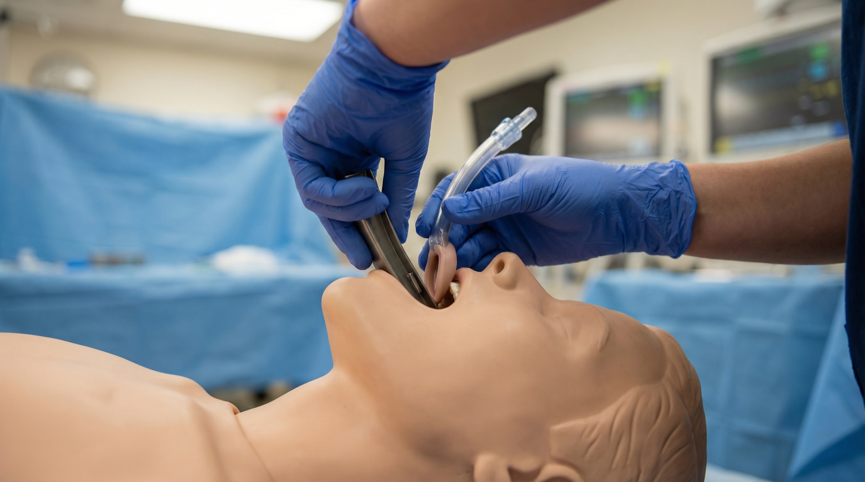
The most common and devastating consequence of cardiac arrest is hypoxic-ischemic brain injury (HIBI). During cardiac arrest, global cerebral ischemia begins within seconds. Irreversible neuronal death can begin within minutes if circulation is not restored. Even after ROSC, a cascade of secondary injury mechanisms — reperfusion injury, excitotoxicity, free radical production, mitochondrial dysfunction, and cerebral edema — continues to damage brain tissue for hours to days. This post-resuscitation injury complex is why the period immediately following ROSC is so clinically critical, and why understanding the immediate post-cardiac arrest care algorithm is foundational for every ACLS provider.

Clinicians benefit from understanding neurological injury after cardiac arrest as a phased process, with distinct pathophysiology and management priorities at each stage.
The primary neurological injury begins the moment cardiac output ceases. Global cerebral ischemia leads to rapid depletion of glucose and ATP, failure of ion pumps, and cellular swelling. Neurons in areas with the highest metabolic demand — particularly the hippocampus, cortex, basal ganglia, and cerebellum — are most vulnerable. The duration of no-flow and low-flow time (time between arrest and initiation of CPR, and time between CPR and ROSC) is the single strongest determinant of initial neurological injury severity. This is precisely why the quality of CPR during the resuscitation phase is so consequential — high-performance CPR techniques that minimize interruptions and maintain adequate perfusion pressure are directly linked to better neurological outcomes.
The secondary injury phase unfolds over the first 24 to 72 hours after ROSC and is paradoxically triggered by the restoration of circulation. Reperfusion injury involves a burst of reactive oxygen species, activation of inflammatory cascades, and disruption of the blood-brain barrier. Simultaneously, patients are vulnerable to systemic insults that can worsen brain outcomes: hypoxia, hypotension, fever, hyperglycemia, and seizures all compound primary brain injury.
Managing these secondary insults is the central focus of post-cardiac arrest critical care. Key interventions include maintaining oxygen saturation between 94% and 98% (avoiding both hypoxia and hyperoxia), targeting a mean arterial pressure of at least 65 mmHg (or higher in selected patients), and implementing targeted temperature management protocols to attenuate neuroinflammation and reduce secondary brain injury. Fever, defined as a temperature above 37.7°C to 38°C, must be aggressively controlled, as even modest hyperthermia significantly worsens neurological outcomes.
Hyperglycemia following cardiac arrest is common and independently associated with worse neurological outcomes. Elevated blood glucose fuels anaerobic metabolism in ischemic penumbra tissue, exacerbates acidosis, and impairs mitochondrial function. Tight glucose control — targeting blood glucose between 140 and 180 mg/dL — is a cornerstone of post-arrest neurological protection. For a detailed clinical breakdown of evidence-based glucose targets, the comprehensive guide to glucose control in post-cardiac arrest care offers practical protocols and clinical context.
One of the most challenging aspects of post-cardiac arrest care is answering the question families ask most urgently: "Will my loved one wake up? Will they be themselves again?" Neurological prognostication — the process of predicting meaningful recovery versus persistent vegetative state or death — is a high-stakes clinical endeavor that demands both rigor and humility.
A critical principle endorsed by the Neurocritical Care Society and the American Heart Association is that neuroprognostication should be deferred. Current guidelines recommend waiting at least 72 hours after ROSC — and at least 72 hours after completion of rewarming in patients who received targeted temperature management — before drawing conclusions about neurological prognosis. The rationale is straightforward: sedatives, paralytics, hypothermia, and metabolic derangements can all mimic neurological devastation. Premature withdrawal of life-sustaining therapy based on early examination findings has led to preventable deaths in patients who would have achieved meaningful recovery.
Research published as part of the guidelines for neuroprognostication in comatose adult survivors of cardiac arrest emphasizes that no single predictor is sufficiently reliable in isolation — a multimodal approach combining clinical examination, electrophysiology, neuroimaging, and biomarkers provides the most accurate prognostic picture.
Modern neuroprognostication integrates multiple modalities, each adding incremental predictive value:
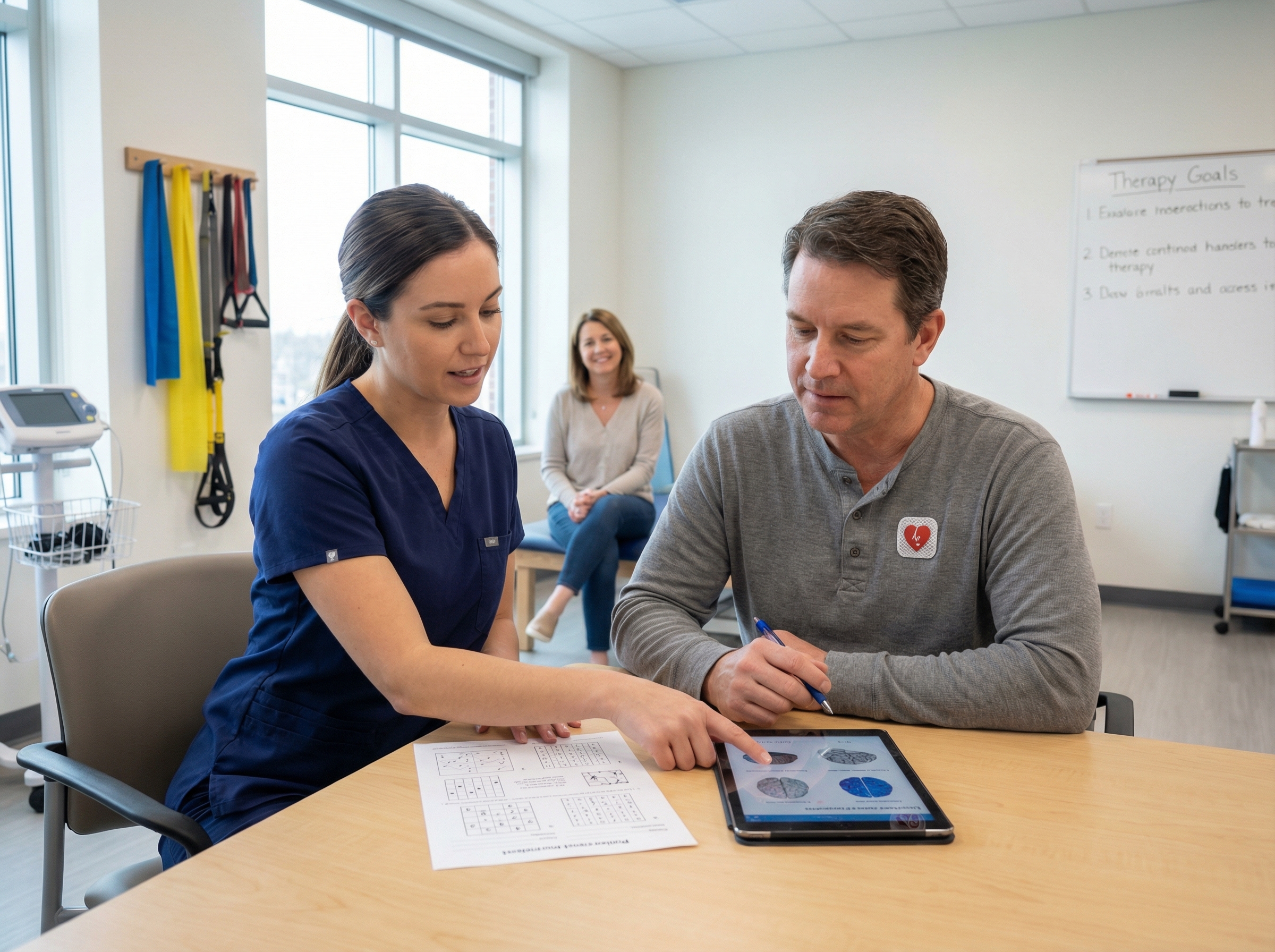
For survivors who emerge from coma and demonstrate meaningful neurological function, recovery follows a trajectory that most families and even many clinicians do not fully appreciate. Neurological recovery after cardiac arrest is not simply a matter of waking up — it is a prolonged, variable process that can span months and sometimes years.
In patients with mild to moderate HIBI, meaningful neurological recovery often begins within the first one to two weeks post-arrest. Patients may progress from coma to a minimally conscious state, then to periods of wakefulness, followed by progressive improvement in cognition, communication, and functional capacity. The speed and extent of this early recovery are shaped by the severity of initial brain injury, pre-arrest neurological status, age, and the quality of acute post-arrest care.
Among those who achieve early neurological recovery, participation in structured rehabilitation programs — beginning in the ICU or step-down unit with early mobilization, speech therapy, and occupational therapy — has been shown to accelerate and enhance recovery. These interventions capitalize on the brain's neuroplasticity: the capacity to reorganize and form new neural connections in response to injury and rehabilitation stimuli. Connecting the resuscitation phase to the recovery phase begins at ROSC — comprehensive post-ROSC care sets the neurological stage for everything that follows.
Even among survivors who appear neurologically intact at hospital discharge, subtle and not-so-subtle cognitive impairments are common. Research published in Frontiers in Aging Neuroscience documents that domains most commonly affected include memory (particularly new learning and recall), attention, processing speed, and executive function. These deficits may not be apparent during brief clinical assessments but can profoundly affect a patient's ability to return to work, manage complex tasks, and maintain relationships.
The psychological burden of cardiac arrest survivorship is equally significant. According to research highlighted by the American Heart Association's cardiac arrest recovery resources, 45 to 50 percent of survivors experience depression, more than a quarter develop post-traumatic stress disorder (PTSD), and up to 61 percent experience clinically significant anxiety. These rates are substantially higher than in the general population and approach those seen in other major trauma survivors. Importantly, these psychological conditions are treatable — early screening and referral to mental health services should be a routine component of post-arrest survivorship care.
For patients who survive to hospital discharge with favorable neurological status, long-term prognosis is more encouraging than many clinicians or patients expect. Long-term survival studies demonstrate that 10-year survival in neurologically intact OHCA survivors ranges from 62% to 64% — a meaningful figure that underscores the value of aggressive resuscitation and post-arrest care in appropriate patients. A significant proportion of survivors with good neurological outcomes return to independent living, work, and social activities.
However, long-term outcomes are not uniformly positive. Survivors face elevated rates of recurrent cardiac events, cognitive decline, and functional limitations that can worsen over time. A systematic review and meta-analysis published through JAMA Cardiology found that while early survival rates have improved, long-term survivors continue to face elevated mortality compared to age-matched controls. Underlying cardiac disease — the most common cause of arrest — requires ongoing management to prevent recurrence.
For patients with severe neurological injury at discharge, the picture is more complex. Some patients in minimally conscious states continue to show slow, incremental improvement over months to years. Families and clinicians should be cautious about drawing permanent conclusions too early. Post-discharge rehabilitation in specialized neurorehabilitation programs can support continued recovery even in patients with significant deficits. Understanding the upstream events that precipitate cardiac arrest — including recognizing warning signs and risk factors — reinforces the value of preventing cardiac catastrophes before they occur.
Understanding neurological recovery after cardiac arrest is not purely an academic exercise — it shapes how clinicians communicate with patients and families, how they structure follow-up care, and how they coordinate the multidisciplinary team required for comprehensive survivorship support.
Families of post-arrest patients often receive prognostic information that is either overly pessimistic (leading to premature withdrawal of care) or falsely optimistic (setting unrealistic expectations for recovery). Best practice involves honest, staged conversations that acknowledge uncertainty, set realistic expectations, and revisit prognosis as new information becomes available. Neurological recovery after cardiac arrest is probabilistic — not deterministic — and families deserve guidance that reflects this nuance.
Clinicians who understand the science of post-arrest neurological recovery are better equipped to have these conversations with confidence and compassion. This knowledge is reinforced through formal ACLS training, which covers the full arc of post-cardiac arrest care. Reflecting on real-life ACLS cases and their outcomes helps providers appreciate how clinical decisions translate into patient stories — including stories of remarkable neurological recovery that might have seemed impossible in the acute phase.
A scientific statement from the American Heart Association on sudden cardiac arrest survivorship recommends that all cardiac arrest survivors receive structured follow-up addressing four key domains: cardiac, neurological, psychological, and social. This survivorship framework acknowledges that recovery extends far beyond hospital discharge and that coordinated, multidisciplinary support significantly improves long-term outcomes.
Key components of an effective survivorship program include:
The outcomes described above — favorable neurological recovery, meaningful long-term survival, return to independent function — are not distributed randomly across patients and hospitals. They are shaped, in part, by the quality of care delivered at every step: the speed and effectiveness of initial resuscitation, the rigor of post-arrest critical care, the sophistication of neuroprognostication, and the comprehensiveness of survivorship follow-up. Clinician knowledge and preparedness at each stage matters.
This is why ACLS certification is not merely a credentialing formality — it is a clinical investment that directly affects patient neurological outcomes. ACLS-trained providers who understand the post-cardiac arrest care algorithm, the rationale for temperature management, the importance of avoiding secondary insults, and the principles of neuroprognostication are better equipped to make the decisions that protect the recovering brain.
The connections between resuscitation quality and neurological outcomes are also well illustrated by advances in extracorporeal CPR. For patients with refractory cardiac arrest, emerging evidence supports the use of ECMO-based resuscitation strategies in selected centers. Understanding the role of extracorporeal CPR technology in advanced resuscitation illustrates how the science of cardiac arrest care continues to evolve toward better neurological preservation.
Life after cardiac arrest is increasingly survivable — and increasingly recoverable — with advances in resuscitation science, post-arrest critical care, and neurological rehabilitation. The picture of neurological recovery after cardiac arrest is nuanced: shaped by the severity of initial ischemic injury, the quality of resuscitation, the rigor of post-arrest management, and the comprehensiveness of long-term survivorship support. Most recovery of cognitive function occurs within the first three months, but improvement can continue for a year or longer. Psychological sequelae are common and treatable. Long-term survival in neurologically intact survivors is better than many expect.
For the clinicians who care for cardiac arrest patients at every stage of this journey — from the first compression to the last outpatient rehabilitation appointment — deep clinical knowledge is itself a form of neuroprotection. Staying current with ACLS guidelines, understanding the science of post-arrest brain injury, and knowing how to communicate prognosis with both honesty and hope are the skills that make the difference between a survival story and a neurological recovery story.
If you are ready to deepen your knowledge of post-cardiac arrest care and sharpen the clinical skills that protect neurological outcomes, Affordable ACLS offers comprehensive, ER physician-developed ACLS certification and recertification courses starting at just $89 — 100% online, self-paced, with immediate certification upon passing. The knowledge you build today directly supports the recovery of every patient whose heart starts beating again under your care.
.jpg)