ACLS Blogs

Lidocaine vs Amiodarone in ACLS: When to Choose This Alternative Antiarrhythmic for Shock-Refractory VF/VT

The Antiarrhythmic Dilemma at the Bedside

You are two shocks into a resuscitation. The monitor still shows coarse ventricular fibrillation. CPR is ongoing, epinephrine has been pushed, and your team is ready for the next defibrillation attempt. Someone hands you the drug box. Amiodarone or lidocaine? For many providers, this decision gets made on autopilot — amiodarone first, always. But the evidence tells a more nuanced story, and understanding it can sharpen your decision-making when it matters most.


Both amiodarone and lidocaine are listed as acceptable antiarrhythmics in current ACLS algorithms for shock-refractory ventricular fibrillation (VF) and pulseless ventricular tachycardia (pVT). Neither has definitively proven superiority in overall survival to discharge. Yet there are clinical scenarios where lidocaine may be the smarter choice — or at minimum, an equally rational one. This article breaks down the pharmacology, the landmark trial data, and the practical situations where you might reach for lidocaine instead of amiodarone.

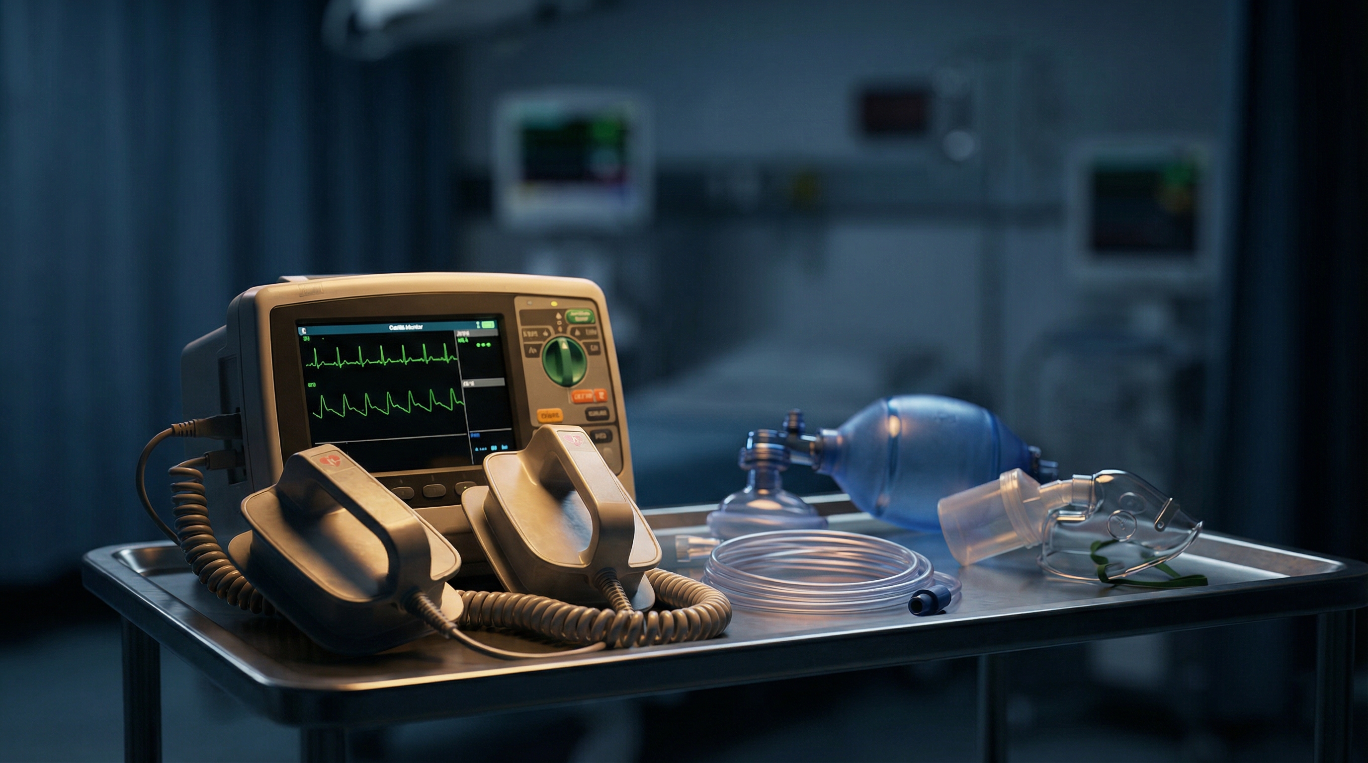
Emergency team reviewing ventricular fibrillation rhythm on cardiac monitor during resuscitation


Understanding the Pharmacology: Two Different Mechanisms

Before comparing clinical outcomes, it helps to understand how these drugs actually work, because their mechanisms of action shape when each one is most appropriate.


Amiodarone: The Multi-Channel Blocker

Amiodarone is a Class III antiarrhythmic agent, but that classification understates its complexity. It blocks potassium channels (prolonging the action potential duration and refractory period), sodium channels, calcium channels, and has noncompetitive beta-blocking properties. This broad, multi-channel activity is part of why it has historically been favored — it attacks arrhythmias through several pathways simultaneously. Its onset of action after IV bolus is typically within minutes for acute VF suppression, though its full antiarrhythmic effect develops over a longer period. The drug is highly lipid-soluble, extensively tissue-bound, and has a half-life measured in weeks. In the acute resuscitation setting, however, you are working with a single bolus dose, so those pharmacokinetic complexities matter less than the immediate electrophysiologic effect.


Lidocaine: The Targeted Sodium Channel Blocker

Lidocaine is a Class Ib antiarrhythmic that works by blocking fast sodium channels, particularly in ischemic or depolarized myocardial tissue. This selectivity is actually one of its underappreciated strengths: lidocaine preferentially binds to sodium channels in a use-dependent and voltage-dependent fashion, meaning it works hardest where the arrhythmia is most active — in rapidly firing, depolarized tissue. It has a short half-life (approximately 90 minutes), rapid onset, and a relatively predictable pharmacokinetic profile. For decades it was the go-to antiarrhythmic in cardiac arrest before amiodarone displaced it following earlier trials. Understanding this targeted mechanism helps explain why lidocaine may actually be advantageous in specific clinical settings, particularly when the underlying cause of VF is ischemic in origin.


The ALPS Trial: What the Evidence Actually Shows

The most important piece of evidence in this debate is the Resuscitation Outcomes Consortium ALPS trial (ROC-ALPS), published in the New England Journal of Medicine in 2016. This randomized, double-blind, placebo-controlled trial enrolled 3,026 patients with out-of-hospital cardiac arrest due to shock-refractory VF or pulseless VT across 10 North American sites.


The results were striking in their equivalence. Survival to hospital discharge was 24.4% in the amiodarone group, 23.7% in the lidocaine group, and 21.0% in the placebo group. Neither amiodarone nor lidocaine produced a statistically significant improvement over placebo in the primary outcome of survival to hospital discharge or favorable neurological outcome. This finding challenged years of clinical dogma and raised an important question: if neither drug clearly beats placebo, is there still a meaningful difference between the two?


A critically important secondary finding from ALPS involved timing. When drugs were administered early — within approximately 10 minutes of arrest — both amiodarone and lidocaine showed notably better performance. In the early-administration subgroup, amiodarone survival to hospital admission reached 62.0% versus 48.5% for placebo (p=0.001). The average time to drug administration in the trial was 19 minutes, which likely diluted the overall benefit. The message for clinical practice: antiarrhythmics work best when given early, and the timing of drug delivery may matter more than which drug you choose. The full ALPS trial data is published in the New England Journal of Medicine and remains essential reading for any ACLS provider.


Shock-Refractory VF/VT: When Do You Give Antiarrhythmics?

Both the AHA and ILCOR guidelines recommend considering antiarrhythmic therapy after the third defibrillation attempt in shock-refractory VF or pulseless VT. The current 2018 AHA focused update on antiarrhythmic drug use during cardiac arrest classifies both amiodarone and lidocaine as reasonable options (Class IIb recommendation), acknowledging comparable clinical utility based on the available evidence.


The standard ACLS dosing for these drugs in cardiac arrest is well-established. For amiodarone: 300 mg IV/IO bolus as the first dose, with a potential repeat dose of 150 mg IV/IO. For lidocaine: 1 to 1.5 mg/kg IV/IO as the first dose, with repeat doses of 0.5 to 0.75 mg/kg IV/IO every 5-10 minutes, up to a maximum total dose of 3 mg/kg. Knowing these doses cold is part of what separates a confident code response from a hesitant one, and it is exactly the kind of clinical knowledge reinforced in ACLS medication cheat sheets covering dosages, routes, and indications.


When to Choose Lidocaine: Five Clinical Scenarios

Given equivalent evidence, the choice between lidocaine and amiodarone in shock-refractory VF/pVT should be driven by clinical context, drug availability, and patient-specific factors. Here are five situations where lidocaine deserves serious consideration as your first antiarrhythmic choice.


1. Ischemia-Driven Ventricular Fibrillation

Lidocaine's use-dependent sodium channel blockade gives it a theoretical — and clinically plausible — advantage in arrhythmias driven by acute myocardial ischemia. The drug preferentially suppresses abnormal automaticity and re-entry in ischemic tissue. When your patient's VF is clearly related to an acute STEMI or high-suspicion ACS (ST changes on the pre-arrest ECG, known CAD, troponin elevation), lidocaine's mechanism is well-suited to the underlying pathophysiology. This is one reason some cardiologists and emergency physicians prefer lidocaine when the arrest is clearly ischemic in etiology rather than a primary electrical disturbance.


2. Amiodarone Contraindicated or Unavailable

Amiodarone should be avoided in patients with known hypersensitivity to amiodarone or iodine, and its use requires caution in patients with severe sinus node dysfunction or second- and third-degree AV block without pacemaker backup (in patients who survive to spontaneous circulation). More practically, amiodarone is sometimes unavailable due to drug shortages — a real-world concern that has affected hospital formularies intermittently. In settings where amiodarone is not in the crash cart or has been substituted, lidocaine is not a fallback option; it is a fully guideline-supported first-line alternative. Wide complex tachycardia management including amiodarone and cardioversion covers additional context on when cardioversion may be preferred over pharmacological therapy.


3. The Post-Cardiac Surgery Patient

Ventricular fibrillation occurring after cardiac surgery represents a unique clinical population. Patients in this setting often have underlying electrolyte disturbances, temporary pacing wires, and freshly reperfused myocardium. Some institutional protocols and expert opinion favor lidocaine in the post-cardiac surgery arrest due to its rapid offset and more predictable pharmacokinetics. The goal is suppressing ischemia-mediated reentry without the prolonged QT effects or hemodynamic complications that amiodarone can occasionally produce. While the evidence base is less robust for this specific subgroup, the pharmacological rationale for lidocaine is sound.


4. Cocaine-Induced Ventricular Arrhythmias

Cocaine toxicity deserves special mention. Cocaine blocks fast sodium channels (mimicking a Class I antiarrhythmic effect) and causes significant sympathomimetic excess, predisposing to wide-complex tachycardias and VF. Amiodarone is theoretically less ideal here because its sodium channel blocking properties may be unpredictable in the setting of already-blocked channels. Lidocaine, as a direct sodium channel blocker with a well-understood mechanism, has been used in cocaine-associated arrhythmias and may offer a more mechanistically rational approach. Cocaine overdose and wide complex tachycardia management covers this specific presentation in further detail.


5. Concern About QT Prolongation After ROSC

Both drugs can prolong the QT interval, but amiodarone's prolonged tissue half-life means QT effects can persist for days to weeks after a single loading dose. If your patient achieves ROSC and you are concerned about ongoing QT prolongation — for instance, in a patient who was already on QT-prolonging medications, or who had pre-existing long QT syndrome, or in whom torsades de pointes is a concern — lidocaine's shorter duration of action offers a pharmacokinetic advantage. Its effects clear within hours, allowing better titration and easier management of post-ROSC arrhythmias without compounding a QT burden that could trigger new problems.


Amiodarone vs. Lidocaine: A Direct Clinical Comparison

To synthesize the evidence, it helps to look at these two drugs side by side across the dimensions that matter most in an acute resuscitation setting.


  • Mechanism of action: Amiodarone is a multi-channel blocker (Class III with I, II, and IV properties). Lidocaine is a targeted Class Ib sodium channel blocker with use-dependent selectivity for depolarized/ischemic tissue.
  • Survival to discharge: Equivalent based on ALPS trial data (24.4% amiodarone vs. 23.7% lidocaine vs. 21.0% placebo; no statistically significant difference between active drugs).
  • ROSC rates: Some earlier data suggested amiodarone improved shock-to-ROSC conversion rates compared to lidocaine in specific populations, but this was not consistently replicated. A 2024 systematic review and meta-analysis found no significant difference in ROSC, short-term survival, or neurological outcomes between the two agents.
  • Half-life and clearance: Amiodarone has a half-life of weeks; lidocaine has a half-life of approximately 90 minutes. Lidocaine clears faster and is more pharmacokinetically predictable post-ROSC.
  • QT prolongation: Both prolong QT, but amiodarone's effect persists far longer due to tissue distribution. Lidocaine carries lower risk for sustained QT prolongation post-resuscitation.
  • Availability and cost: Lidocaine is generally less expensive and has a more stable supply chain. Both are available as IV formulations compatible with IV/IO administration.
  • Ischemia-specificity: Lidocaine has theoretical pharmacological advantage in arrhythmias driven by acute myocardial ischemia due to use-dependent sodium channel blockade in depolarized tissue.
  • Dosing complexity: Lidocaine dosing is weight-based (1-1.5 mg/kg), while amiodarone is given as a fixed bolus (300 mg). Some providers find amiodarone's fixed dosing simpler under pressure, though either can be administered confidently with preparation.


Where Antiarrhythmics Fit in the ACLS Cardiac Arrest Algorithm

It is worth stepping back to situate antiarrhythmic therapy within the broader cardiac arrest algorithm. The foundation of resuscitation for shockable rhythms remains high-quality CPR and prompt defibrillation. Antiarrhythmics are an adjunct — they are administered to patients who have not responded to multiple defibrillation attempts, and they should never come at the expense of minimizing interruptions to compressions or delaying a shock.


In the adult cardiac arrest algorithm, antiarrhythmics are introduced in the third cycle of resuscitation — after the second shock, with epinephrine already on board. At this point, whether you choose amiodarone or lidocaine, the administration technique matters enormously: give the drug as a rapid IV/IO bolus, followed immediately by a 20 mL fluid flush, and continue CPR without delay. Time from drug push to defibrillation shock should be minimized. Understanding ACLS medication timing and drug delivery windows helps providers understand how to sequence interventions effectively during a resuscitation.


Remember that epinephrine (1 mg IV/IO every 3-5 minutes) remains the primary vasopressor in cardiac arrest algorithms for both shockable and non-shockable rhythms. Antiarrhythmics are specific to shockable rhythms (VF and pVT) and are not indicated for asystole or pulseless electrical activity. Epinephrine versus vasopressin in cardiac arrest explores the vasopressor evidence base in further depth for providers interested in the full pharmacological picture of ACLS resuscitation.


After ROSC: Continuing Antiarrhythmic Therapy

Once spontaneous circulation is restored, the question of whether to continue antiarrhythmic therapy shifts to a different evidence framework. If VF or pVT recurs in the post-ROSC period, re-dosing or initiating a continuous infusion may be appropriate. For amiodarone, a maintenance infusion of 1 mg/min for 6 hours followed by 0.5 mg/min for 18 hours is standard if a bolus was given during the arrest. For lidocaine, a maintenance infusion of 1 to 4 mg/min can be initiated after successful resuscitation when the drug was used as the antiarrhythmic during arrest.


Post-ROSC care is a complex and demanding phase. Targeted temperature management, hemodynamic optimization, and urgent cardiac catheterization for suspected STEMI all take priority. The antiarrhythmic you used during arrest informs but does not fully dictate post-ROSC management. Post-ROSC care: what happens after the heart starts beating again provides a comprehensive overview of the immediate post-resuscitation phase for providers preparing to manage these critically ill patients.


Special Populations and Considerations

Several patient populations require modified thinking about antiarrhythmic selection in VF/pVT arrest.


Pediatric Cardiac Arrest

In pediatric cardiac arrest with shockable rhythms, amiodarone and lidocaine are both listed as options in current PALS guidelines, with dosing adjusted for weight. Pediatric VF is less common than in adults and more often secondary to respiratory failure or hypoxia, but when it does occur in the context of structural heart disease or channelopathies, the same antiarrhythmic principles apply. Weight-based dosing becomes even more critical, and the complexity of pediatric resuscitation underscores the value of comprehensive PALS training.


Cardiac Arrest in Pregnancy

Both amiodarone and lidocaine cross the placenta. Amiodarone carries known fetal risks including hypothyroidism, bradycardia, and premature birth when used chronically; however, in the acute setting of maternal cardiac arrest, the priority is maternal resuscitation. Lidocaine also crosses the placenta but with generally more predictable short-term effects. When VF occurs in a pregnant patient, the same ACLS algorithm applies — modified for the lateral uterine displacement — and the choice of antiarrhythmic should follow the same evidence-based rationale discussed throughout this article.


Pre-Existing Structural Heart Disease

Patients with known cardiomyopathy, prior myocardial infarction, or reduced ejection fraction may already be on long-term oral amiodarone. In these patients, presenting to arrest with amiodarone already on board, additional IV amiodarone loading during resuscitation carries uncertain pharmacokinetic implications given the drug's massive tissue distribution volume. Some clinicians in this scenario would transition to lidocaine as the IV antiarrhythmic rather than stacking amiodarone doses, though definitive evidence for this practice is limited.


Practical Tips for the Code Room

Knowing the pharmacology and evidence is necessary but not sufficient. Here are practical considerations for antiarrhythmic decision-making during an active resuscitation.

Healthcare provider preparing antiarrhythmic medication at crash cart during cardiac arrest response


  • Know your doses cold. Hesitation over drug dosing during a code costs precious seconds. Whether your institution defaults to amiodarone or lidocaine, be able to recall the dose immediately under pressure: amiodarone 300 mg IV/IO, lidocaine 1-1.5 mg/kg IV/IO.
  • Check your cart and formulary before the code. Drug shortages are real. Know what is available in your code cart so you are not surprised mid-resuscitation.
  • Communicate your choice clearly. As team leader, call out both the drug and the dose: "Push amiodarone 300 milligrams IV push now." Closed-loop communication prevents errors and keeps the team synchronized.
  • Document the time of drug administration. The ALPS trial's timing subgroup data reinforces that early administration matters. Tracking when drugs were pushed helps post-event debriefing and quality improvement.
  • Never delay a shock for a drug push. If the patient is ready to be shocked, shock first. Drugs are given during the CPR phase immediately following defibrillation. The rhythm — not the drug order — drives timing.
  • Let the clinical context guide your choice. If the arrest is clearly ischemic, cocaine-related, or the patient has concerns about prolonged QT post-ROSC, lean toward lidocaine. If no specific contraindication or preference exists, either drug is acceptable.


Mastering Antiarrhythmics Through ACLS Training

The nuances of antiarrhythmic selection — knowing not just what the algorithm says, but why it says it, and when to thoughtfully deviate — represent the difference between algorithmic ACLS and genuinely expert resuscitation. This kind of clinical depth is what separates providers who have truly internalized the science from those who are merely memorizing steps.


That depth of understanding is what Affordable ACLS is built to deliver. Founded by board-certified emergency medicine physicians with over 20 years of combined clinical experience, our 100% online, self-paced ACLS certification course goes beyond rote algorithm recitation to explain the pharmacological rationale behind every drug decision, the evidence basis for every recommendation, and the practical judgment that separates a good resuscitation from a great one. At just $99 for certification and $89 for recertification, it is designed for working clinicians who need rigorous, flexible training without disrupting their schedule or draining their budget.


Whether you are an RN, physician, PA, NP, paramedic, or respiratory therapist, understanding shockable rhythms including ventricular tachycardia and fibrillation at a mechanistic level makes you a better provider the moment you step into a code. Our course covers ACLS medications, algorithms, rhythm interpretation, and team dynamics — everything you need to act with confidence when the monitor alarms and seconds count.


The Bottom Line: Evidence-Based Flexibility

The question of lidocaine versus amiodarone for shock-refractory VF/pVT does not have a single correct answer, and that is actually a strength of current evidence-based practice rather than a weakness. The ALPS trial demonstrated that both drugs are reasonable, that early administration matters more than drug selection, and that neither drug is so clearly superior as to demand exclusive use. The 2018 AHA focused update reflects this by listing both as Class IIb options without a preferred hierarchy.


What this means in practice is that the clinician who understands both drugs — their mechanisms, their pharmacokinetics, their clinical niches — is better equipped than one who has simply memorized "amiodarone first." When VF is clearly ischemic, choose lidocaine. When amiodarone is contraindicated or unavailable, choose lidocaine. When QT prolongation post-ROSC is a concern, consider lidocaine. When no specific factor tips the balance, either drug is defensible and guideline-supported.


Mastering this kind of nuanced pharmacological judgment is precisely what distinguishes the excellent ACLS provider from the competent one. If you are preparing for ACLS certification or recertification — or simply want to sharpen your clinical knowledge in resuscitation pharmacology — Affordable ACLS offers the rigorous, clinically grounded training you need, on your schedule, at a price that respects your investment in your career.


ACLS Blogs

Lidocaine vs Amiodarone in ACLS: When to Choose This Alternative Antiarrhythmic for Shock-Refractory VF/VT

The Antiarrhythmic Dilemma at the Bedside

You are two shocks into a resuscitation. The monitor still shows coarse ventricular fibrillation. CPR is ongoing, epinephrine has been pushed, and your team is ready for the next defibrillation attempt. Someone hands you the drug box. Amiodarone or lidocaine? For many providers, this decision gets made on autopilot — amiodarone first, always. But the evidence tells a more nuanced story, and understanding it can sharpen your decision-making when it matters most.


Both amiodarone and lidocaine are listed as acceptable antiarrhythmics in current ACLS algorithms for shock-refractory ventricular fibrillation (VF) and pulseless ventricular tachycardia (pVT). Neither has definitively proven superiority in overall survival to discharge. Yet there are clinical scenarios where lidocaine may be the smarter choice — or at minimum, an equally rational one. This article breaks down the pharmacology, the landmark trial data, and the practical situations where you might reach for lidocaine instead of amiodarone.

Emergency team reviewing ventricular fibrillation rhythm on cardiac monitor during resuscitation


Understanding the Pharmacology: Two Different Mechanisms

Before comparing clinical outcomes, it helps to understand how these drugs actually work, because their mechanisms of action shape when each one is most appropriate.


Amiodarone: The Multi-Channel Blocker

Amiodarone is a Class III antiarrhythmic agent, but that classification understates its complexity. It blocks potassium channels (prolonging the action potential duration and refractory period), sodium channels, calcium channels, and has noncompetitive beta-blocking properties. This broad, multi-channel activity is part of why it has historically been favored — it attacks arrhythmias through several pathways simultaneously. Its onset of action after IV bolus is typically within minutes for acute VF suppression, though its full antiarrhythmic effect develops over a longer period. The drug is highly lipid-soluble, extensively tissue-bound, and has a half-life measured in weeks. In the acute resuscitation setting, however, you are working with a single bolus dose, so those pharmacokinetic complexities matter less than the immediate electrophysiologic effect.


Lidocaine: The Targeted Sodium Channel Blocker

Lidocaine is a Class Ib antiarrhythmic that works by blocking fast sodium channels, particularly in ischemic or depolarized myocardial tissue. This selectivity is actually one of its underappreciated strengths: lidocaine preferentially binds to sodium channels in a use-dependent and voltage-dependent fashion, meaning it works hardest where the arrhythmia is most active — in rapidly firing, depolarized tissue. It has a short half-life (approximately 90 minutes), rapid onset, and a relatively predictable pharmacokinetic profile. For decades it was the go-to antiarrhythmic in cardiac arrest before amiodarone displaced it following earlier trials. Understanding this targeted mechanism helps explain why lidocaine may actually be advantageous in specific clinical settings, particularly when the underlying cause of VF is ischemic in origin.


The ALPS Trial: What the Evidence Actually Shows

The most important piece of evidence in this debate is the Resuscitation Outcomes Consortium ALPS trial (ROC-ALPS), published in the New England Journal of Medicine in 2016. This randomized, double-blind, placebo-controlled trial enrolled 3,026 patients with out-of-hospital cardiac arrest due to shock-refractory VF or pulseless VT across 10 North American sites.


The results were striking in their equivalence. Survival to hospital discharge was 24.4% in the amiodarone group, 23.7% in the lidocaine group, and 21.0% in the placebo group. Neither amiodarone nor lidocaine produced a statistically significant improvement over placebo in the primary outcome of survival to hospital discharge or favorable neurological outcome. This finding challenged years of clinical dogma and raised an important question: if neither drug clearly beats placebo, is there still a meaningful difference between the two?


A critically important secondary finding from ALPS involved timing. When drugs were administered early — within approximately 10 minutes of arrest — both amiodarone and lidocaine showed notably better performance. In the early-administration subgroup, amiodarone survival to hospital admission reached 62.0% versus 48.5% for placebo (p=0.001). The average time to drug administration in the trial was 19 minutes, which likely diluted the overall benefit. The message for clinical practice: antiarrhythmics work best when given early, and the timing of drug delivery may matter more than which drug you choose. The full ALPS trial data is published in the New England Journal of Medicine and remains essential reading for any ACLS provider.


Shock-Refractory VF/VT: When Do You Give Antiarrhythmics?

Both the AHA and ILCOR guidelines recommend considering antiarrhythmic therapy after the third defibrillation attempt in shock-refractory VF or pulseless VT. The current 2018 AHA focused update on antiarrhythmic drug use during cardiac arrest classifies both amiodarone and lidocaine as reasonable options (Class IIb recommendation), acknowledging comparable clinical utility based on the available evidence.


The standard ACLS dosing for these drugs in cardiac arrest is well-established. For amiodarone: 300 mg IV/IO bolus as the first dose, with a potential repeat dose of 150 mg IV/IO. For lidocaine: 1 to 1.5 mg/kg IV/IO as the first dose, with repeat doses of 0.5 to 0.75 mg/kg IV/IO every 5-10 minutes, up to a maximum total dose of 3 mg/kg. Knowing these doses cold is part of what separates a confident code response from a hesitant one, and it is exactly the kind of clinical knowledge reinforced in ACLS medication cheat sheets covering dosages, routes, and indications.


When to Choose Lidocaine: Five Clinical Scenarios

Given equivalent evidence, the choice between lidocaine and amiodarone in shock-refractory VF/pVT should be driven by clinical context, drug availability, and patient-specific factors. Here are five situations where lidocaine deserves serious consideration as your first antiarrhythmic choice.


1. Ischemia-Driven Ventricular Fibrillation

Lidocaine's use-dependent sodium channel blockade gives it a theoretical — and clinically plausible — advantage in arrhythmias driven by acute myocardial ischemia. The drug preferentially suppresses abnormal automaticity and re-entry in ischemic tissue. When your patient's VF is clearly related to an acute STEMI or high-suspicion ACS (ST changes on the pre-arrest ECG, known CAD, troponin elevation), lidocaine's mechanism is well-suited to the underlying pathophysiology. This is one reason some cardiologists and emergency physicians prefer lidocaine when the arrest is clearly ischemic in etiology rather than a primary electrical disturbance.


2. Amiodarone Contraindicated or Unavailable

Amiodarone should be avoided in patients with known hypersensitivity to amiodarone or iodine, and its use requires caution in patients with severe sinus node dysfunction or second- and third-degree AV block without pacemaker backup (in patients who survive to spontaneous circulation). More practically, amiodarone is sometimes unavailable due to drug shortages — a real-world concern that has affected hospital formularies intermittently. In settings where amiodarone is not in the crash cart or has been substituted, lidocaine is not a fallback option; it is a fully guideline-supported first-line alternative. Wide complex tachycardia management including amiodarone and cardioversion covers additional context on when cardioversion may be preferred over pharmacological therapy.


3. The Post-Cardiac Surgery Patient

Ventricular fibrillation occurring after cardiac surgery represents a unique clinical population. Patients in this setting often have underlying electrolyte disturbances, temporary pacing wires, and freshly reperfused myocardium. Some institutional protocols and expert opinion favor lidocaine in the post-cardiac surgery arrest due to its rapid offset and more predictable pharmacokinetics. The goal is suppressing ischemia-mediated reentry without the prolonged QT effects or hemodynamic complications that amiodarone can occasionally produce. While the evidence base is less robust for this specific subgroup, the pharmacological rationale for lidocaine is sound.


4. Cocaine-Induced Ventricular Arrhythmias

Cocaine toxicity deserves special mention. Cocaine blocks fast sodium channels (mimicking a Class I antiarrhythmic effect) and causes significant sympathomimetic excess, predisposing to wide-complex tachycardias and VF. Amiodarone is theoretically less ideal here because its sodium channel blocking properties may be unpredictable in the setting of already-blocked channels. Lidocaine, as a direct sodium channel blocker with a well-understood mechanism, has been used in cocaine-associated arrhythmias and may offer a more mechanistically rational approach. Cocaine overdose and wide complex tachycardia management covers this specific presentation in further detail.


5. Concern About QT Prolongation After ROSC

Both drugs can prolong the QT interval, but amiodarone's prolonged tissue half-life means QT effects can persist for days to weeks after a single loading dose. If your patient achieves ROSC and you are concerned about ongoing QT prolongation — for instance, in a patient who was already on QT-prolonging medications, or who had pre-existing long QT syndrome, or in whom torsades de pointes is a concern — lidocaine's shorter duration of action offers a pharmacokinetic advantage. Its effects clear within hours, allowing better titration and easier management of post-ROSC arrhythmias without compounding a QT burden that could trigger new problems.


Amiodarone vs. Lidocaine: A Direct Clinical Comparison

To synthesize the evidence, it helps to look at these two drugs side by side across the dimensions that matter most in an acute resuscitation setting.


  • Mechanism of action: Amiodarone is a multi-channel blocker (Class III with I, II, and IV properties). Lidocaine is a targeted Class Ib sodium channel blocker with use-dependent selectivity for depolarized/ischemic tissue.
  • Survival to discharge: Equivalent based on ALPS trial data (24.4% amiodarone vs. 23.7% lidocaine vs. 21.0% placebo; no statistically significant difference between active drugs).
  • ROSC rates: Some earlier data suggested amiodarone improved shock-to-ROSC conversion rates compared to lidocaine in specific populations, but this was not consistently replicated. A 2024 systematic review and meta-analysis found no significant difference in ROSC, short-term survival, or neurological outcomes between the two agents.
  • Half-life and clearance: Amiodarone has a half-life of weeks; lidocaine has a half-life of approximately 90 minutes. Lidocaine clears faster and is more pharmacokinetically predictable post-ROSC.
  • QT prolongation: Both prolong QT, but amiodarone's effect persists far longer due to tissue distribution. Lidocaine carries lower risk for sustained QT prolongation post-resuscitation.
  • Availability and cost: Lidocaine is generally less expensive and has a more stable supply chain. Both are available as IV formulations compatible with IV/IO administration.
  • Ischemia-specificity: Lidocaine has theoretical pharmacological advantage in arrhythmias driven by acute myocardial ischemia due to use-dependent sodium channel blockade in depolarized tissue.
  • Dosing complexity: Lidocaine dosing is weight-based (1-1.5 mg/kg), while amiodarone is given as a fixed bolus (300 mg). Some providers find amiodarone's fixed dosing simpler under pressure, though either can be administered confidently with preparation.


Where Antiarrhythmics Fit in the ACLS Cardiac Arrest Algorithm

It is worth stepping back to situate antiarrhythmic therapy within the broader cardiac arrest algorithm. The foundation of resuscitation for shockable rhythms remains high-quality CPR and prompt defibrillation. Antiarrhythmics are an adjunct — they are administered to patients who have not responded to multiple defibrillation attempts, and they should never come at the expense of minimizing interruptions to compressions or delaying a shock.


In the adult cardiac arrest algorithm, antiarrhythmics are introduced in the third cycle of resuscitation — after the second shock, with epinephrine already on board. At this point, whether you choose amiodarone or lidocaine, the administration technique matters enormously: give the drug as a rapid IV/IO bolus, followed immediately by a 20 mL fluid flush, and continue CPR without delay. Time from drug push to defibrillation shock should be minimized. Understanding ACLS medication timing and drug delivery windows helps providers understand how to sequence interventions effectively during a resuscitation.


Remember that epinephrine (1 mg IV/IO every 3-5 minutes) remains the primary vasopressor in cardiac arrest algorithms for both shockable and non-shockable rhythms. Antiarrhythmics are specific to shockable rhythms (VF and pVT) and are not indicated for asystole or pulseless electrical activity. Epinephrine versus vasopressin in cardiac arrest explores the vasopressor evidence base in further depth for providers interested in the full pharmacological picture of ACLS resuscitation.


After ROSC: Continuing Antiarrhythmic Therapy

Once spontaneous circulation is restored, the question of whether to continue antiarrhythmic therapy shifts to a different evidence framework. If VF or pVT recurs in the post-ROSC period, re-dosing or initiating a continuous infusion may be appropriate. For amiodarone, a maintenance infusion of 1 mg/min for 6 hours followed by 0.5 mg/min for 18 hours is standard if a bolus was given during the arrest. For lidocaine, a maintenance infusion of 1 to 4 mg/min can be initiated after successful resuscitation when the drug was used as the antiarrhythmic during arrest.


Post-ROSC care is a complex and demanding phase. Targeted temperature management, hemodynamic optimization, and urgent cardiac catheterization for suspected STEMI all take priority. The antiarrhythmic you used during arrest informs but does not fully dictate post-ROSC management. Post-ROSC care: what happens after the heart starts beating again provides a comprehensive overview of the immediate post-resuscitation phase for providers preparing to manage these critically ill patients.


Special Populations and Considerations

Several patient populations require modified thinking about antiarrhythmic selection in VF/pVT arrest.


Pediatric Cardiac Arrest

In pediatric cardiac arrest with shockable rhythms, amiodarone and lidocaine are both listed as options in current PALS guidelines, with dosing adjusted for weight. Pediatric VF is less common than in adults and more often secondary to respiratory failure or hypoxia, but when it does occur in the context of structural heart disease or channelopathies, the same antiarrhythmic principles apply. Weight-based dosing becomes even more critical, and the complexity of pediatric resuscitation underscores the value of comprehensive PALS training.


Cardiac Arrest in Pregnancy

Both amiodarone and lidocaine cross the placenta. Amiodarone carries known fetal risks including hypothyroidism, bradycardia, and premature birth when used chronically; however, in the acute setting of maternal cardiac arrest, the priority is maternal resuscitation. Lidocaine also crosses the placenta but with generally more predictable short-term effects. When VF occurs in a pregnant patient, the same ACLS algorithm applies — modified for the lateral uterine displacement — and the choice of antiarrhythmic should follow the same evidence-based rationale discussed throughout this article.


Pre-Existing Structural Heart Disease

Patients with known cardiomyopathy, prior myocardial infarction, or reduced ejection fraction may already be on long-term oral amiodarone. In these patients, presenting to arrest with amiodarone already on board, additional IV amiodarone loading during resuscitation carries uncertain pharmacokinetic implications given the drug's massive tissue distribution volume. Some clinicians in this scenario would transition to lidocaine as the IV antiarrhythmic rather than stacking amiodarone doses, though definitive evidence for this practice is limited.


Practical Tips for the Code Room

Knowing the pharmacology and evidence is necessary but not sufficient. Here are practical considerations for antiarrhythmic decision-making during an active resuscitation.

Healthcare provider preparing antiarrhythmic medication at crash cart during cardiac arrest response


  • Know your doses cold. Hesitation over drug dosing during a code costs precious seconds. Whether your institution defaults to amiodarone or lidocaine, be able to recall the dose immediately under pressure: amiodarone 300 mg IV/IO, lidocaine 1-1.5 mg/kg IV/IO.
  • Check your cart and formulary before the code. Drug shortages are real. Know what is available in your code cart so you are not surprised mid-resuscitation.
  • Communicate your choice clearly. As team leader, call out both the drug and the dose: "Push amiodarone 300 milligrams IV push now." Closed-loop communication prevents errors and keeps the team synchronized.
  • Document the time of drug administration. The ALPS trial's timing subgroup data reinforces that early administration matters. Tracking when drugs were pushed helps post-event debriefing and quality improvement.
  • Never delay a shock for a drug push. If the patient is ready to be shocked, shock first. Drugs are given during the CPR phase immediately following defibrillation. The rhythm — not the drug order — drives timing.
  • Let the clinical context guide your choice. If the arrest is clearly ischemic, cocaine-related, or the patient has concerns about prolonged QT post-ROSC, lean toward lidocaine. If no specific contraindication or preference exists, either drug is acceptable.


Mastering Antiarrhythmics Through ACLS Training

The nuances of antiarrhythmic selection — knowing not just what the algorithm says, but why it says it, and when to thoughtfully deviate — represent the difference between algorithmic ACLS and genuinely expert resuscitation. This kind of clinical depth is what separates providers who have truly internalized the science from those who are merely memorizing steps.


That depth of understanding is what Affordable ACLS is built to deliver. Founded by board-certified emergency medicine physicians with over 20 years of combined clinical experience, our 100% online, self-paced ACLS certification course goes beyond rote algorithm recitation to explain the pharmacological rationale behind every drug decision, the evidence basis for every recommendation, and the practical judgment that separates a good resuscitation from a great one. At just $99 for certification and $89 for recertification, it is designed for working clinicians who need rigorous, flexible training without disrupting their schedule or draining their budget.


Whether you are an RN, physician, PA, NP, paramedic, or respiratory therapist, understanding shockable rhythms including ventricular tachycardia and fibrillation at a mechanistic level makes you a better provider the moment you step into a code. Our course covers ACLS medications, algorithms, rhythm interpretation, and team dynamics — everything you need to act with confidence when the monitor alarms and seconds count.


The Bottom Line: Evidence-Based Flexibility

The question of lidocaine versus amiodarone for shock-refractory VF/pVT does not have a single correct answer, and that is actually a strength of current evidence-based practice rather than a weakness. The ALPS trial demonstrated that both drugs are reasonable, that early administration matters more than drug selection, and that neither drug is so clearly superior as to demand exclusive use. The 2018 AHA focused update reflects this by listing both as Class IIb options without a preferred hierarchy.


What this means in practice is that the clinician who understands both drugs — their mechanisms, their pharmacokinetics, their clinical niches — is better equipped than one who has simply memorized "amiodarone first." When VF is clearly ischemic, choose lidocaine. When amiodarone is contraindicated or unavailable, choose lidocaine. When QT prolongation post-ROSC is a concern, consider lidocaine. When no specific factor tips the balance, either drug is defensible and guideline-supported.


Mastering this kind of nuanced pharmacological judgment is precisely what distinguishes the excellent ACLS provider from the competent one. If you are preparing for ACLS certification or recertification — or simply want to sharpen your clinical knowledge in resuscitation pharmacology — Affordable ACLS offers the rigorous, clinically grounded training you need, on your schedule, at a price that respects your investment in your career.


blogs

More articles for you

all articles

Start Today!

Get certified at your own pace, on your schedule, from the comfort of your home.