Endotracheal Aspiration (ETA) is one of the most critical interventions in modern critical care medicine. This essential procedure involves the systematic removal of respiratory secretions from intubated patients through specialized suction techniques, serving as a cornerstone of airway management in intensive care units worldwide.
When you encounter patients who cannot effectively clear their own respiratory secretions due to sedation, neurological impairment, or mechanical ventilation, ETA becomes their lifeline. The procedure directly addresses the fundamental challenge of maintaining open airways in critically ill individuals who have lost their natural protective mechanisms.
The significance of ETA extends beyond simple secretion removal. This intervention prevents life-threatening complications including airway obstruction, ventilator-associated pneumonia, and respiratory failure. Healthcare professionals rely on this technique to maintain optimal ventilation and oxygenation in patients with tracheostomy care needs or those requiring prolonged mechanical ventilation.
You will discover the comprehensive aspects of endotracheal aspiration throughout this guide, including:
Mastering ETA techniques directly impacts patient survival rates and recovery trajectories in critical care environments, making this knowledge indispensable for healthcare professionals working with intubated patients.
For healthcare professionals seeking to enhance their skills in managing such critical situations, enrolling in advanced courses like the ACLS & BLS Recertification Bundle for Groups can be immensely beneficial. These courses provide comprehensive training that includes first-time recertification for both ACLS and BLS, unlimited retakes if necessary at no charge, and downloadable provider cards immediately after completion.
Moreover, understanding the risk assessment and complication management strategies associated with ETA is crucial for optimal patient outcomes. Such knowledge can significantly improve the quality of care provided to intubated patients.
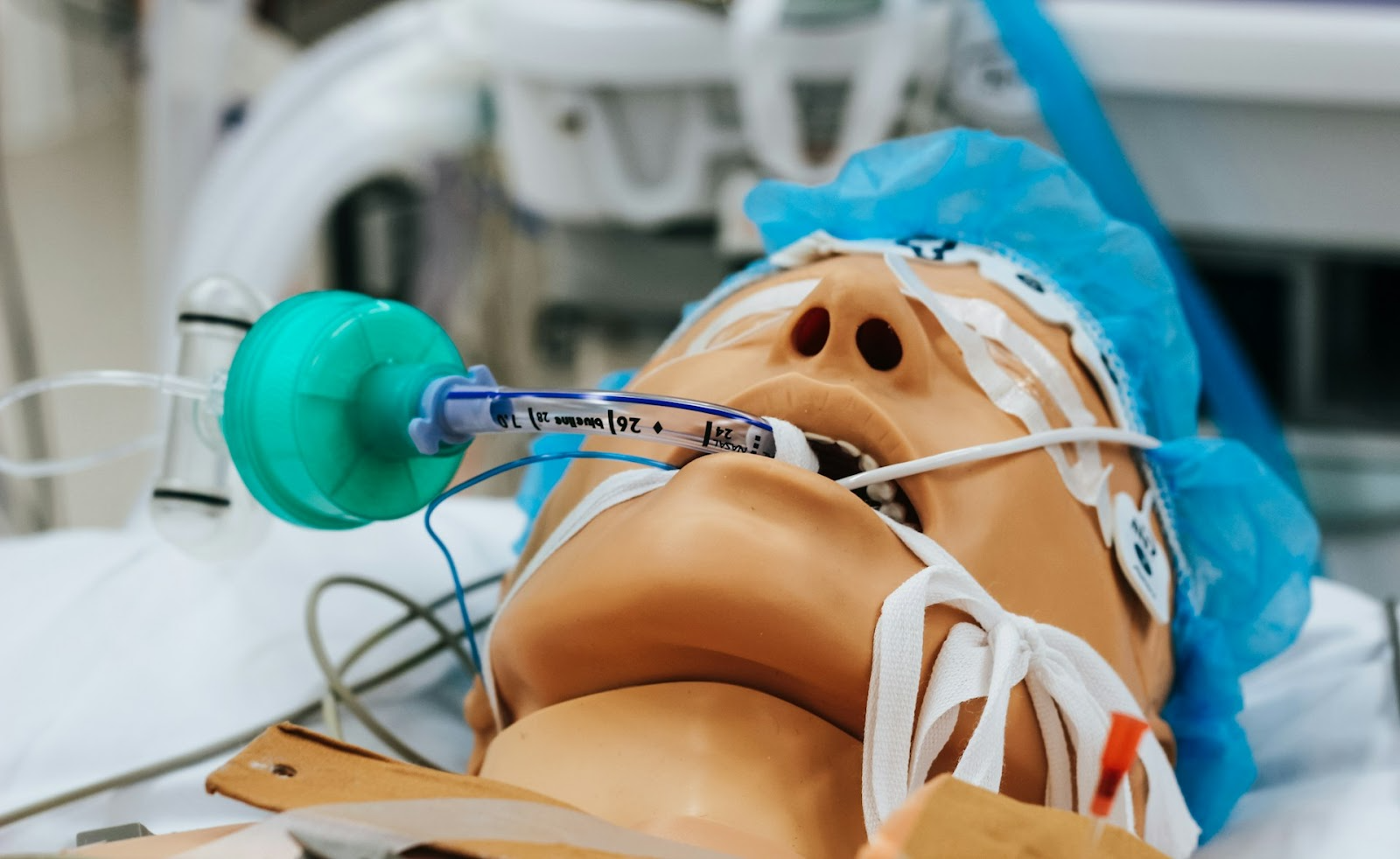
Endotracheal aspiration (ETA) is a basic procedure that involves using a catheter to remove excess fluid or mucus from the airways of patients who are on a ventilator. This technique is performed by inserting a sterile suction tube through an artificial airway (such as an endotracheal tube or tracheostomy tube) in order to extract any buildup of mucus, blood, or other substances that may be affecting the patient's ability to breathe.
The procedure uses special suction catheters that are designed to safely navigate through endotracheal tubes or tracheostomy tubes. Healthcare providers connect the catheter to a vacuum source, which creates controlled negative pressure and pulls secretions from the patient's respiratory system. The flexibility of the catheter allows it to reach different areas within the bronchial tree, ensuring thorough removal of secretions.
During the procedure, you insert the catheter without applying suction until it reaches the desired depth. Once positioned, you activate the suction while slowly withdrawing the catheter in a rotating motion. This technique maximizes secretion removal while minimizing trauma to the delicate respiratory tissues.
Secretion Clearance serves as the most common reason for performing ETA. Patients who have difficulty coughing, produce excessive amounts of mucus, or have thick secretions may require mechanical assistance to keep their airways clear.
Preventing Airway Obstruction becomes critical when secretions accumulate to levels that impede ventilation. You'll recognize this need through decreased oxygen saturation, increased peak airway pressures, or visible secretions in the artificial airway.
Improving Patient Outcomes extends beyond immediate respiratory relief. Regular ETA procedures help prevent ventilator-associated pneumonia, reduce the risk of atelectasis (collapse of lung tissue), and maintain optimal gas exchange. The procedure also allows healthcare providers to evaluate secretion characteristics such as color, consistency, and volume—information that can aid in diagnosing respiratory conditions and infections.
The timing and frequency of ETA procedures depend on individual patient needs rather than predetermined schedules. This approach ensures personalized care that adapts to changing clinical situations.
For those involved in critical care procedures like ETA, it's important to have a solid understanding of advanced cardiovascular life support (ACLS) protocols. Being familiar with ACLS algorithms can greatly improve patient outcomes during emergencies. Regular training and recertification courses are essential for healthcare professionals to stay updated on the latest practices and guidelines in ACLS.

To perform endotracheal aspiration (ETA) successfully, it's crucial to have a good grasp of the artificial airways used in this procedure. There are two main types of artificial airways commonly used in critical care settings for ETA: endotracheal tubes and tracheostomy tubes.
The endotracheal tube is the most frequently used artificial airway during emergency intubation and mechanical ventilation. This flexible plastic tube can be inserted through either the mouth or nose and is positioned directly into the trachea during a procedure called endotracheal intubation. By placing the tube in this manner, we create a secure airway that bypasses the upper respiratory tract completely.
Here are some important characteristics of endotracheal tubes:
Unlike endotracheal tubes, which provide temporary airway access, tracheostomy tubes offer a more permanent solution. These tubes are surgically inserted through an opening in the neck (known as a tracheostomy) and enter directly into the trachea, bypassing both the mouth and nose.
Tracheostomy tubes come with several benefits:
Both types of devices play critical roles in maintaining an open airway during medical procedures. However, they serve different purposes based on specific situations:
Understanding these distinctions helps healthcare professionals make informed decisions about which device to use based on individual patient needs.
When it comes to performing ETA procedures effectively, both tube types directly influence suction catheter selection and insertion depth:
By considering these anatomical relationships between various devices involved in ETA, clinicians can enhance their skills further when it comes time for secretion removal interventions.
While understanding anatomy is vital for successful ETAs alone, being familiar with certain critical care concepts also contributes significantly towards improving patient outcomes during such interventions.
For instance:
These additional insights gained from studying broader principles within critical care medicine complement technical skills required during specific procedures like ETA.
In emergencies where every second counts, having proficiency in [basic life support techniques](https://affordableacls.com/lessons/2-solo-provider-adult-bls) becomes invaluable alongside knowledge related specifically towards ETAs themselves.
Such skills include:
By integrating these foundational resuscitation strategies alongside specialized approaches tailored towards managing airways through methods such as endotracheal aspiration , healthcare professionals can optimize chances at successful outcomes even under challenging circumstances .
Respiratory distress is the main reason why we need to do Endotracheal Aspiration (ETA) on Intubated Patients. We can tell when someone is having trouble breathing by looking for signs like they are working harder to breathe, using their neck and chest muscles to help them breathe, or struggling to get air in and out. If a patient on a ventilator is showing these signs, we need to act quickly and suction their airway to remove any blockages that are making it difficult for them to breathe.
Sometimes, the situation may get worse and we might need to do more advanced procedures like the Post Cardiac Arrest Algorithm, especially if the patient is showing severe difficulty breathing that could lead to heart problems.
Ineffective cough mechanisms are another important reason why we need to perform ETA. Patients who are sedated, have brain injuries, or have muscle diseases may not be able to cough effectively and clear their airways. In these cases, we need to suction their airway when we see mucus buildup that would normally be cleared by coughing.
Oxygen saturation levels are also important in deciding when we need to suction. We should continuously monitor the patient's oxygen levels using a pulse oximeter and look out for any drops below their normal range. If we see a decrease of 3-5% in their oxygen saturation, it usually means we need to do ETA right away.
Compromised respiratory function in patients on mechanical ventilation can happen due to different lung diseases or conditions. Patients with chronic obstructive pulmonary disease, pneumonia, or acute respiratory distress syndrome often require regular suctioning to keep their lungs working well. These patients tend to produce a lot of mucus that affects how oxygen and carbon dioxide are exchanged in their lungs and how well the ventilator is synchronized with their breathing.
The timing of when we do ETA depends on how each patient is doing instead of following a set schedule. We need to assess each patient's breathing condition, how much mucus they are producing, and how their oxygen levels are changing over time to figure out the best frequency for suctioning. By focusing on what each patient needs, we can provide effective treatment while avoiding unnecessary procedures that could lead to complications.
In certain situations, such as when a patient has adult tachycardia with a pulse, we may need to change our approach and include other life-saving actions along with ETA.

Performing endotracheal aspiration requires meticulous attention to detail and adherence to established protocols. The procedure begins with comprehensive preparation to ensure patient safety and optimal outcomes.
Personal protective equipment forms the foundation of safe ETA performance. You must don gloves, gowns, eye protection, and masks before approaching the patient. The sterile technique demands that you maintain aseptic conditions throughout the entire procedure to prevent healthcare-associated infections.
Your preparation checklist includes:
The sterile field must remain uncontaminated from start to finish. You handle the suction catheter with sterile gloves, touching only the proximal end while keeping the distal tip sterile until insertion.
Hyperoxygenation serves as a critical safety measure before initiating suctioning. You increase the patient's fraction of inspired oxygen (FiO2) to 100% for 30-60 seconds prior to catheter insertion. This preemptive step builds oxygen reserves in the patient's lungs, creating a buffer against the inevitable oxygen desaturation that occurs during suctioning.
The hyperoxygenation process involves:
Catheter size selection directly impacts both procedure effectiveness and patient safety. The catheter diameter should not exceed half the internal diameter of the endotracheal tube to prevent excessive suction pressure and airway trauma.
Standard sizing recommendations include:
While performing endotracheal aspiration, it's crucial to consider patient positioning carefully. Generally, it is not recommended to move a victim unless there is a direct danger to their life or if it is necessary to provide care. In such cases, assisting an unconscious but breathing patient into the recovery position can help protect their airway and reduce aspiration risk.
During procedures like endotracheal aspiration, it's vital to be aware of potential complications such as a heart attack which may present symptoms like chest tightness, nausea, sweating, shortness of breath, fatigue, pain in the arm or jaw, and pallor. Recognizing these symptoms early can be life-saving; therefore, immediate action such as calling 911 or preparing to start CPR may be required while also administering appropriate medication like aspirin if advised (source).
Endotracheal aspiration (ETA) is a crucial procedure in critical care that provides immediate treatment and important diagnostic information for patients who are intubated. The main purpose of this procedure is to keep the airway open by removing secretions, which allows for better ventilation.
Respiratory secretions can be a major problem for patients on mechanical ventilation. These secretions can quickly block the airways, leading to breathing difficulties and ineffective gas exchange. ETA directly addresses this issue by:
The benefits of ETA go beyond just clearing secretions. Regularly performing this procedure can also prevent atelectasis, which is the collapse of lung areas caused by blocked airways. You can see evidence of this improvement through better chest X-ray results and increased ventilator compliance measurements after successful suctioning.
Ventilator-associated pneumonia (VAP) is a serious complication that can occur in intubated patients, with high mortality rates among those affected. ETA samples provide important diagnostic information that helps identify the specific bacteria causing VAP and guide treatment decisions.
The diagnostic process involves analyzing the aspirated secretions using culture techniques to identify bacterial growth patterns associated with VAP development. Common bacteria encountered through ETA sampling include:
The combination of therapeutic benefits from secretion clearance and diagnostic insights gained from identifying pathogens makes ETA a valuable intervention in critical care management. By addressing both airway obstruction and potential infections simultaneously, healthcare providers can optimize patient outcomes during mechanical ventilation.
Endotracheal Aspiration (ETA) of Intubated Patients carries inherent risks that healthcare providers must carefully manage to ensure patient safety. Understanding these potential complications enables you to implement preventive measures and respond appropriately when adverse events occur.
Bronchoconstriction represents one of the most immediate risks during suctioning procedures. The mechanical stimulation of the airway can trigger bronchospasm, particularly in patients with reactive airway disease or asthma. You'll notice this complication through sudden increases in peak airway pressures and decreased oxygen saturation levels.
Tracheal mucosa injury occurs when inappropriate technique or excessive suction pressure damages the delicate respiratory epithelium. This trauma can lead to:
The suctioning process can precipitate serious physiological disturbances. Hypoxia develops rapidly if you exceed the recommended 10-second suction duration or fail to provide adequate pre-oxygenation. Cardiac arrhythmias may result from hypoxia, vagal stimulation, or the stress response to the procedure.
Atelectasis can occur when excessive negative pressure collapses alveoli, particularly in patients with compromised lung compliance. You must balance effective secretion removal with gentle technique to minimize this risk.
Neonatal resuscitation scenarios present unique challenges when performing ETA. Newborns have smaller airways, more compliant chest walls, and limited physiological reserves. You need specialized pediatric-sized catheters and reduced suction pressures to prevent airway trauma.
In such cases, understanding the Pediatric Basic Life Support Algorithm is essential for two-rescuer scenarios. This algorithm outlines crucial steps including scene safety, compressions, ventilation, and AED use which are vital during emergencies involving neonates.
Pediatric patients with tracheostomy tubes require modified approaches due to their anatomical differences and increased sensitivity to pressure changes. Their shorter tracheal length means you must carefully control catheter insertion depth to avoid advancing into the bronchi.
Moreover, if you're regularly working with children, obtaining a PALS certification can equip you with essential skills to handle various emergencies including cardiac arrest or severe allergic reactions which could arise during such procedures.
Premature infants face additional risks including intraventricular hemorrhage from pressure fluctuations and apnea episodes triggered by vagal stimulation during suctioning.
Personal protective equipment (PPE) forms the cornerstone of safe ETA performance. You must don appropriate protective gear before initiating any suctioning procedure to safeguard both yourself and your patients from potential infection control breaches. The essential PPE components include:
Clinical guidelines established by professional organizations provide evidence-based frameworks for optimal ETA performance. The American Association for Respiratory Care (AARC) recommends specific protocols that you should follow consistently:
It's crucial to note that during the procedure, AI is transforming emergency cardiac care by improving diagnosis, treatment precision, and patient outcomes through advanced data analysis and real-time decision support.
You should never reuse suction catheters between patients or even between suctioning attempts on the same patient. This practice significantly reduces cross-contamination risks and maintains the highest standards of infection control in critical care environments.
Moreover, it's essential to enhance your skills continually. Following best study tips tailored for online course takers can help you excel in your studies and succeed in your certification journey. Lastly, familiarizing yourself with ACLS algorithms can simplify emergency care training and improve life-saving skills effectively.
The world of endotracheal aspiration is constantly changing as researchers and clinicians strive to improve this life-saving procedure for better patient outcomes in critical care environments. Current medical research is focused on several promising areas that will shape the future of ETA practice.
Advanced suction systems with automated pressure regulation show potential for reducing complications while maintaining effectiveness. Smart monitoring devices that integrate with ventilators can provide real-time feedback about secretion volume and consistency, helping you make more informed decisions about suctioning frequency.
Research teams are investigating patient-specific protocols based on individual risk factors, underlying conditions, and secretion characteristics. This personalized medicine approach aims to minimize unnecessary procedures while ensuring optimal airway management.
New molecular diagnostic techniques promise faster pathogen identification from ETA samples, potentially reducing the time to appropriate antibiotic therapy for ventilator-associated pneumonia. Point-of-care testing devices could revolutionize how you approach infection management in intubated patients.
Critical Care Nursing education programs increasingly emphasize evidence-based ETA techniques, ensuring the next generation of healthcare providers delivers the highest standard of care.
The dual therapeutic and diagnostic value of endotracheal aspiration makes it an indispensable tool for improving outcomes in ICU patients. However, it's essential to remember that successful outcomes often depend on a well-coordinated effort across various medical disciplines. This is where understanding the adult chain of survival becomes crucial, as it outlines the key steps that need to be taken during a medical emergency.
Moreover, enhancing your skills through resources like BLS recertification or guideline changes can significantly improve your ability to manage critical situations effectively. It's also vital to have a solid grasp of post-resuscitation management, which is a critical phase following any resuscitative effort.
Stay informed about ongoing research developments to enhance your practice and provide the best possible care for your intubated patients.
.jpg)