Ventricular fibrillation is the most treatable cause of cardiac arrest — but only when defibrillation works. For the majority of patients, a well-timed shock converts chaotic electrical activity back into a perfusing rhythm. For a significant subset, however, the fibrillation persists despite repeated attempts. This is refractory ventricular fibrillation, and it represents one of the most challenging scenarios any resuscitation team can face.
Double sequential external defibrillation — commonly abbreviated as DSED — has emerged as a clinically promising technique for exactly these situations. By deploying two defibrillators in rapid sequence and repositioning the shock vector, DSED attempts to reach myocardial tissue that standard anterior-lateral pad placement may fail to depolarize. The evidence base is still maturing, but the landmark DOSE-VF trial published in the New England Journal of Medicine brought this approach into serious clinical conversation, and the 2025 AHA/ILCOR guideline updates acknowledge DSED as a legitimate advanced resuscitation option.
This article breaks down the science behind refractory VF, explains how DSED works mechanistically, walks through the clinical evidence including the DOSE-VF results, and discusses how DSED fits into a comprehensive ACLS approach to refractory shockable rhythms. If you manage cardiac arrests in the ED, the ICU, or in the field, this is knowledge you need.
Refractory ventricular fibrillation is generally defined as persistent VF after three or more failed defibrillation attempts, all delivered with full energy per manufacturer recommendation and accompanied by high-quality CPR. The exact threshold varies slightly by institution and protocol, but the core concept is consistent: standard defibrillation has been tried, has failed, and the rhythm has not converted.
The incidence of refractory VF in out-of-hospital cardiac arrest ranges from roughly 10 to 20 percent of all VF cases, depending on the population studied. In absolute terms, that translates to thousands of patients annually across North America and Europe who fail initial defibrillation attempts. For these patients, the prognosis with standard care has historically been poor. Prolonged ischemia compounds organ damage with each passing minute, and repeated unsuccessful shocks do not contribute to survival.
Several factors are thought to contribute to refractoriness. Myocardial substrate issues — including ischemia, electrolyte abnormalities, acidosis, and poor coronary perfusion — all reduce the likelihood of successful defibrillation. Transthoracic impedance, the resistance that current must overcome to reach the myocardium, is another key variable. If the shock vector does not deliver sufficient current density to a critical mass of ventricular myocardium, defibrillation will fail even at high energy levels. This is where DSED and vector-change strategies enter the picture. To fully understand the underlying condition, our comprehensive guide to ventricular fibrillation covers the full pathophysiology in depth.
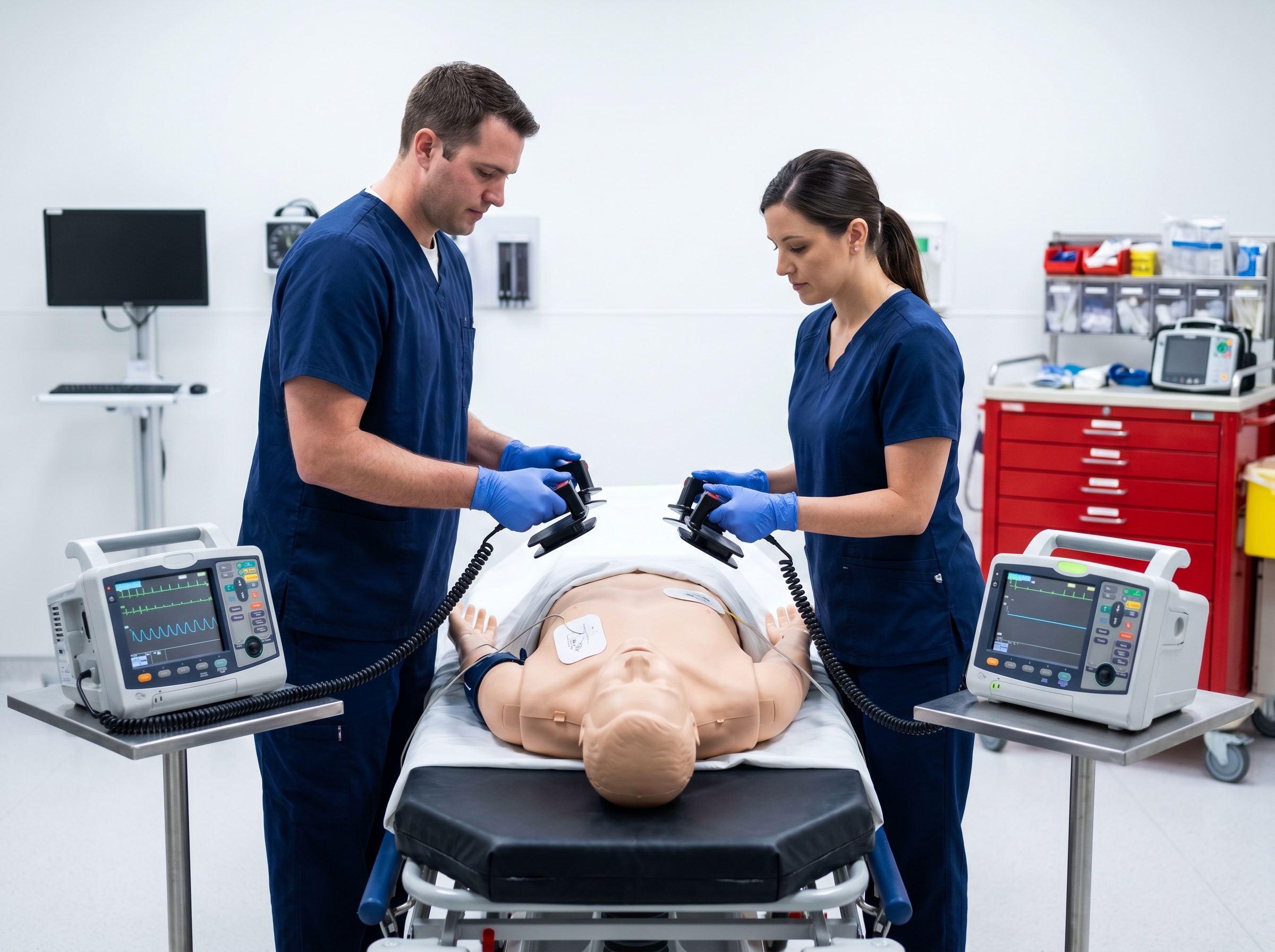
DSED uses two separate defibrillators in rapid sequence, with electrode pads placed in two different configurations. The standard setup involves the first defibrillator with pads in the conventional anterior-lateral position, and the second defibrillator with pads in the anterior-posterior position. When both units fire in close succession — typically within a fraction of a second — the combined electrical vectors create a broader field of depolarization across the ventricle.
The theoretical advantage is straightforward: if the reason standard defibrillation is failing is that a region of the myocardium lies outside the effective depolarization zone of the anterior-lateral vector, adding a second vector from a different axis increases the probability that all critical ventricular tissue receives enough current to terminate fibrillation. Research from the DOSE-VF trial measured transthoracic impedance across defibrillation attempts and found a consistent 10 to 20 percent reduction in impedance during the anterior-posterior shock compared to the prior failed anterior-lateral attempt — suggesting that repositioned pads are delivering energy more efficiently to previously under-served myocardium.
It is important to distinguish DSED from vector-change defibrillation, a related but distinct technique. Vector-change defibrillation simply repositions a single set of pads from anterior-lateral to anterior-posterior placement before delivering the next shock — no second defibrillator is required. Both approaches aim to reach under-depolarized tissue, but DSED adds the additional energy of a second simultaneous or near-simultaneous shock. The DOSE-VF trial studied both strategies against standard defibrillation, making it the only randomized controlled trial to date directly comparing all three approaches.
The DOSE-VF (DOuble SEquential External Defibrillation for Refractory Ventricular Fibrillation) trial was published in the New England Journal of Medicine in 2022 and remains the primary landmark study driving clinical adoption of DSED. The trial enrolled 405 adult patients with out-of-hospital cardiac arrest who remained in VF after initial defibrillation attempts. Participants were randomized to one of three groups: standard defibrillation (136 patients), DSED (125 patients), or vector-change defibrillation (144 patients).
The results were striking. Survival to hospital discharge was 30 percent in the DSED group compared to 13 percent with standard defibrillation — a statistically significant and clinically meaningful difference. The vector-change group also outperformed standard defibrillation, with a discharge survival rate of 24 percent. Rates of VF termination and return of spontaneous circulation (ROSC) were higher in both the DSED and vector-change arms compared to controls. These outcomes represent a near-doubling of survival probability for a patient population that had historically faced extremely poor prognosis.

The neurological outcomes were equally encouraging. Among survivors in the DSED group, favorable neurological function at hospital discharge was significantly more common than in the standard defibrillation group. This is a critical point: it is not enough to achieve ROSC if the patient survives in a persistent vegetative state. The DOSE-VF data suggest that DSED's benefit extends to meaningful long-term survival, not simply return of cardiac activity.
That said, the trial has limitations that must be acknowledged. It was conducted across a specific paramedic system with defined protocols and training requirements. Generalizability to all EMS systems and in-hospital settings requires careful consideration of local training, equipment availability, and coordination capacity. Subsequent meta-analyses and systematic reviews have shown mixed results when incorporating smaller observational studies, underscoring that DSED's benefit appears clearest in well-controlled, protocol-driven environments.
From a practical standpoint, DSED requires preparation that goes beyond having an extra defibrillator available. Successful execution depends on team coordination, equipment compatibility checks, and role clarity before the technique is needed. Improvising DSED during a code in which the team has never practiced the technique is likely to introduce delays and execution errors that negate any potential benefit.
The standard approach requires positioning four electrode pads: anterior-lateral pads from the first defibrillator (right subclavian to left lateral chest) and anterior-posterior pads from the second defibrillator (left anterior precordium to left posterior thorax). Both defibrillators should be charged simultaneously, and the shocks delivered in rapid succession — ideally within one to two seconds of each other. Truly simultaneous firing from two separate devices has not been validated in terms of device safety, and most protocols call for near-simultaneous rather than truly concurrent discharge. Healthcare providers should follow the manufacturer guidance for their specific equipment and any institutional protocol in place.
One critical practice point from the Resuscitation Council UK and emerging expert consensus: once DSED or vector-change defibrillation has been initiated, it should be maintained for all subsequent defibrillation attempts throughout the resuscitation, including after ROSC and in the event of rearrest. Reverting to standard anterior-lateral placement after transitioning to an advanced strategy is not supported by current evidence and may reintroduce the same impedance barriers that caused initial defibrillation failure. For the pharmacology that runs in parallel with these decisions, our comparison of lidocaine versus amiodarone for shock-refractory VF/VT is essential reading for any provider managing refractory rhythms.
DSED does not replace ACLS pharmacology for refractory VF — it complements it. Current ACLS protocols call for amiodarone 300 mg IV/IO after the third failed shock, with a second dose of 150 mg available if the rhythm persists. Lidocaine remains an acceptable alternative, particularly in settings where amiodarone is unavailable or contraindicated. These antiarrhythmic drugs work through different mechanisms than defibrillation: while shocks aim to terminate fibrillation through mass depolarization, antiarrhythmics modify the electrophysiological substrate to make the myocardium less prone to re-fibrillation.
The optimal timing of DSED relative to antiarrhythmic administration is still a matter of ongoing research. In the DOSE-VF trial, antiarrhythmic use was permitted but not standardized across arms, making it difficult to disentangle the independent contribution of each intervention. Practically speaking, most clinicians initiate antiarrhythmic therapy in parallel with advanced defibrillation strategies rather than treating them as sequential steps. The 2025 AHA/ILCOR guidelines acknowledge DSED as a legitimate option for refractory shockable rhythms, but do not yet provide specific position statements on timing relative to drug administration.
Epinephrine 1 mg IV/IO every 3 to 5 minutes remains part of the standard algorithm for all shockable rhythms. Its role in refractory VF is unchanged by DSED — vasopressor support for coronary and cerebral perfusion pressure continues to run in the background regardless of defibrillation strategy. Providers should also be vigilant about reversible causes (the H's and T's) that may be driving continued VF, including hyperkalemia, acidosis, and tension pneumothorax, as correcting the underlying cause may allow defibrillation to succeed where it otherwise would not. Our article on amiodarone and cardioversion in wide complex tachycardia provides further context on antiarrhythmic decision-making during shockable rhythms.
An important caveat for providers considering DSED: as of this writing, defibrillator technology in the United States and Europe is not specifically approved for use in a DSED shock sequence. Off-label use of medical devices is a common and legally recognized practice, but it does carry institutional and medicolegal implications that providers should be aware of. Implementing DSED as a formal protocol at your institution should involve medical director oversight, and ideally a written institutional protocol that documents the evidence basis and training requirements.
From an equipment standpoint, most hospitals and many advanced EMS systems already have more than one defibrillator available. The marginal equipment cost of implementing DSED is therefore often modest. The primary investment is in training — ensuring that resuscitation team members understand the indication, technique, and coordination requirements before they face a refractory VF situation in real time. Mock code practice is an ideal venue for incorporating DSED into team muscle memory. For those looking to strengthen their overall cardiac arrest management foundation, our guide to high-performance CPR team strategies covers the broader systems approach that DSED fits within.
DSED sits in the middle of what many resuscitation experts now conceptualize as an escalating continuum of interventions for refractory VF. Standard defibrillation is the first-line approach. Vector-change defibrillation is a low-resource next step, requiring only pad repositioning. DSED adds a second defibrillator. At the far end of the continuum sits extracorporeal CPR (ECPR) — the use of venoarterial ECMO to provide mechanical circulatory support during continued resuscitation efforts.
ECPR has shown survival benefit in select patients with refractory arrest, particularly younger patients with shockable rhythms and short no-flow times. However, ECPR requires specialized infrastructure, a trained perfusion team, and rapid cannulation capability — resources that are simply not available in most clinical settings. DSED, by contrast, is logistically accessible to any team with two defibrillators and appropriate training. This positions DSED as a critical intermediate step: more aggressive than pad repositioning alone, but dramatically more accessible than ECPR. For a deeper look at where ECPR fits into the refractory arrest algorithm, our article on ECPR and advanced resuscitation technology is a valuable companion read.
The emerging consensus framework, as reflected in the American Heart Association's 2025 scientific priorities report on DSED, suggests that the decision to escalate should be driven by patient factors (witnessed arrest, initial shockable rhythm, short no-flow time, younger age), system resources, and elapsed resuscitation time. Patients who meet favorable criteria for ECPR should not be anchored to DSED if ECPR is available and cannulation can be accomplished rapidly. Conversely, DSED should not be withheld from patients who are ECPR-ineligible simply because it has not yet received a Class I guideline recommendation.
When DSED achieves ROSC, the post-resuscitation care priorities are identical to those following standard defibrillation: hemodynamic optimization, targeted temperature management consideration, urgent coronary angiography in appropriate candidates, and neurological prognostication. The mechanism by which DSED terminates fibrillation does not change the downstream pathophysiology of post-cardiac arrest syndrome, and post-ROSC care protocols should proceed without modification.
One practical consideration: with two defibrillators and four sets of pads in place, the transition to post-arrest monitoring requires careful pad removal and electrode placement for standard 12-lead ECG acquisition and continuous monitoring. Teams should designate responsibility for defibrillator management during the transition phase to avoid confusion about which device is providing rhythm monitoring and which is available for rearrest. The risk of rearrest in the first hours after ROSC is non-trivial, and having a clear defibrillation plan ready before it is needed is essential. For the full scope of what follows successful resuscitation, our post-ROSC care guide covers the critical management decisions in detail.
Point-of-care ultrasound (POCUS) plays an increasingly important role in both the intra-arrest and post-arrest phases. During resuscitation, POCUS can identify reversible causes — cardiac tamponade, massive pulmonary embolism, severe hypovolemia — that might explain defibrillation resistance and guide additional intervention. After ROSC, echocardiographic assessment of left ventricular function, wall motion, and filling status directly informs vasopressor and fluid management decisions. Understanding how POCUS integrates with ACLS decision-making is covered comprehensively in our POCUS in cardiac arrest article.
Despite the promise of the DOSE-VF trial, significant knowledge gaps remain. The AHA convened a multistakeholder thinktank in 2025 specifically to identify scientific priorities for DSED research, reflecting both the technique's clinical importance and the recognized limitations of the existing evidence base. Key unanswered questions include: What is the optimal energy level for each shock in the DSED sequence? Should the anterior-lateral shock fire first, or should the anterior-posterior shock lead? Is there an additive benefit to combining DSED with antiarrhythmic pretreatment versus administering the drugs after the DSED attempt?
Operational implementation research is also needed. A 2025 systematic review published in Healthcare identified significant barriers to DSED implementation in real-world settings, including training gaps, equipment access delays, and lack of standardized protocols. The review found that even at institutions where DSED is theoretically available, execution failures — wrong pad placement, delayed second shock, confusion about which defibrillator to charge — can eliminate the technique's benefit. Addressing these system-level barriers through simulation training, standardized protocols, and equipment pre-positioning is as important as the underlying science.
The question of in-hospital versus out-of-hospital applicability also remains largely unexplored. The DOSE-VF trial was an out-of-hospital study. In the hospital setting, the team composition, communication dynamics, and equipment logistics differ substantially. In-hospital DSED likely requires different protocol design, and dedicated research in this population is needed before strong recommendations can be made. A network meta-analysis examining defibrillation strategies for refractory VF further highlights the complexity of comparing outcomes across different study designs and patient populations, reinforcing the need for well-powered multicenter trials.
DSED is not a technique that providers stumble into successfully during a high-stakes resuscitation without prior preparation. Like every advanced ACLS skill, its effective application depends on foundational training, team rehearsal, and a mental model built through deliberate study. Understanding the pathophysiology of refractory VF, the mechanism of action of different defibrillation vectors, the pharmacology of antiarrhythmic agents, and the decision points for escalating to ECPR all form the cognitive scaffolding that allows providers to make fast, accurate decisions when a patient is in refractory VF.
This is precisely why ACLS certification and recertification matter — not as checkbox exercises, but as genuine opportunities to update clinical knowledge and reinforce the decision frameworks that determine outcomes. The Affordable ACLS curriculum is built on current AHA and ILCOR guidelines, including the 2025 updates that acknowledge advanced defibrillation strategies for refractory shockable rhythms. Courses are developed by actively practicing, Board Certified Emergency Medicine physicians who understand the clinical realities of managing refractory cardiac arrest from the inside.
With ACLS certification available online for $99 and recertification for $89 — completable in one to two hours at your own pace, on any device — there is no practical barrier to staying current. Unlimited retakes at no extra cost mean you can review materials as many times as necessary to feel genuinely confident, not just credentialed. A money-back guarantee removes any financial risk from the decision to train online. For those specifically managing antiarrhythmic decisions during refractory arrest, our guide to ACLS medication timing and drug delivery windows is a must-read companion resource for providers who want to integrate pharmacology with advanced defibrillation strategies effectively.
Double sequential external defibrillation has moved from the category of experimental curiosity to evidence-informed option in a remarkably short period. The DOSE-VF trial demonstrated survival rates that nearly doubled compared to standard defibrillation in refractory VF, with favorable neurological outcomes among survivors. Subsequent research has complicated the picture somewhat, revealing implementation barriers and calling for larger, multicenter trials — but the signal of benefit, particularly in well-trained, protocol-driven environments, is real.
For providers managing cardiac arrests, the practical takeaway is this: refractory VF is not a terminal declaration. It is a clinical decision point. Vector-change defibrillation requires minimal additional resources and should be the immediate next step when standard defibrillation fails. DSED offers an additional energy vector with growing evidence support and deserves a place in the institutional protocols and training programs of any facility where cardiac arrests are managed. ECPR remains available for the subset of patients who meet eligibility criteria and where infrastructure supports it.
Each of these options requires that providers have the knowledge and training to recognize when to escalate, how to execute the technique, and how to integrate it with the rest of the ACLS algorithm. The science is advancing rapidly — and staying current through evidence-based ACLS training is how individual clinicians keep pace with what the evidence now supports. Visit affordableacls.com to explore certification and recertification options built by EM physicians who understand exactly what clinicians face when a patient is in refractory VF and standard care is not working.
.jpg)